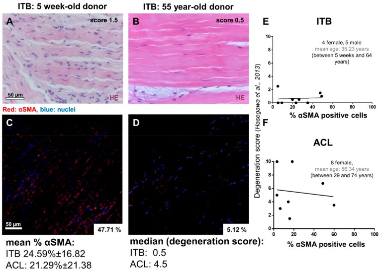
Figure 2.
Representative ITB samples stained with hematoxylin eosin (HE) and immunolabeled for α-smooth muscle actin (αSMA). (A,B) A representative HE staining of ITB tissue of a young and aged donor. (C,D) αSMA-staining of the same samples. No significant correlation could be shown between the stage of degeneration and the percentage of αSMA expressing cells for ITB (E) (spearman: r = 0.149, p = 0.701) and ACL (F) (spearman: r = −0.096, p = 0.806) tissue samples. Scale bars: 50 µm. ITB: iliotibial band, ACL: anterior cruciate ligament.

