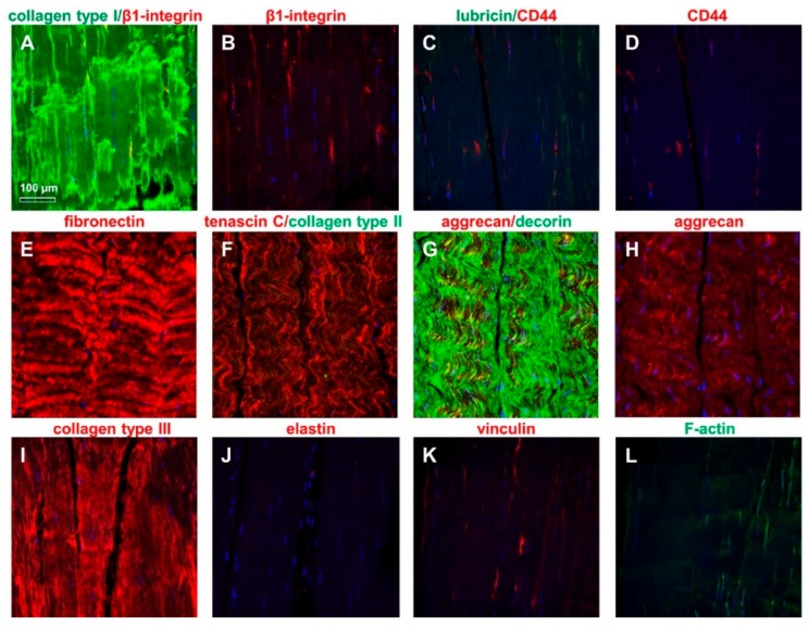
Figure 4.
The immunolabeling of the extracellular matrix (ECM) and cytoplasmatic markers in ITB tissue: Collagen type I (green) and β1-integrin [(A) merged, (B) β1-integrin (red)], lubricin (green)/CD44 (red) [(C) merged, (D) CD44], fibronectin [(E) red], tenascin C (red)/collagen type II (green) (F), aggrecan/decorin [(G) merged, (H) aggrecan], collagen type III [(I) red], elastin [(J) red], vinculin [(K) red], and F-actin [(L) green]. The cell nuclei were counterstained using 4’,6-diamidino-2-phenylindole (DAPI). Scale bar: 100 µm.

