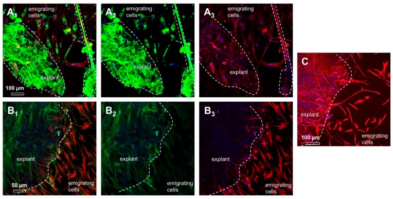
Figure 6.
The expression profile in ITB explants and emigrating cells. (A) The main ligament extracellular matrix (ECM) protein collagen type I and the corresponding cell ECM adhesion receptor β1-integrin are shown (A1: merged, A2: collagen type I (green), and A3: β1-integrin (red)). Dashed line: The explant and, on the right side, a thick bundle of collagen fibers are shown. (B) F-actin filament organization and synthesis of vascular endothelial growth factor A (VEGFA) in explants cultured for 5 weeks and emigrating cells (B1: merged, B2: F-actin (green), and B3: VEGFA (red)). (C) The mesenchymal intermediate filament vimentin (red). Scale bars: Figure 6A,C: 100 µm and Figure 6B: 50 µm.

