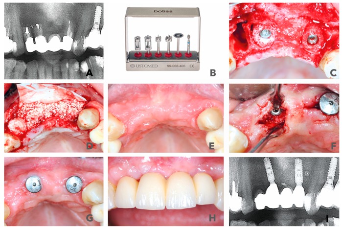
Figure 2.
(A) Radiological display of the bone defect in frontal maxilla #11-22. (B) maxgraft bonering surgical kit. (C) Bone augmentation with two cylindrical FDBAs and fixation with dental implants. (D) Augmentation site contoured with bovine bone substiute and covered with collagen membrane. (E) and (F) Six-month follow-up of contouring and re-entry to place gingiva formers. (G,H) Integration of prosthetic restoration 6 weeks after re-entry and 7.5 months after initial surgery. (I) Thirty-six-month radiological follow-up showed stable bone in augmented area.

