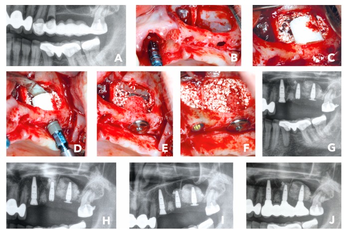
Figure 4.
(A) Initial situation: Radiological image of bridge retained second quadrant with hopeless teeth #23 and #24. (B) External sinus floor elavation and implant placement #23. (C) Placement of a 7-mm bone ring block #27 and bovine bone substitute. (D) Implant placement. (E) Crestal placement of a fication screw to secure the bone graft and implant. (F) Implant placement in #25 and sinus cavity filled with bovine bone substitute. (G) Post-op x-ray. (H) Radiographical control 6 months after surgery. (I) Radiographical control 1 week later when the implants were uncovered and prosthodontic impression made. (J) Radiographical image of final prosthetic restoration 6.5 months after initial surgery.

