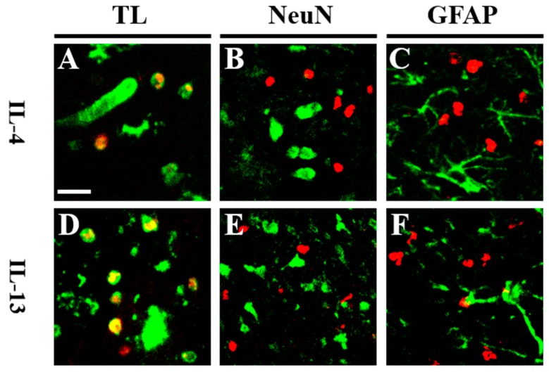
Figure 3.
pKr-2-induced IL-4 and IL-13 are co-localized within activated microglia/macrophages in vivo. (A–F) Animals receiving a unilateral injection of pKr-2 into cerebral cortex were sacrificed 1 day later, brains were removed, and coronal sections (40 μm) were prepared for immunohistochemical staining. Fluorescence images of (A,D) Tomato Lectin (green) for microglia/macrophages and IL-4 (A, red) or IL-13 (D, red), (B,E) NeuN (green) for neurons and IL-4 (B, red) or IL-13 (E, red), and (C,F) glial fibrillary acidic protein (GFAP) (green) for astrocytes and IL-4 (C, red), or IL-13 (F, red). Each image was captured from the similar cortical area and merged (yellow). Scale bar: 25 μm. n = 4 to 6. TL: tomato lectin.

