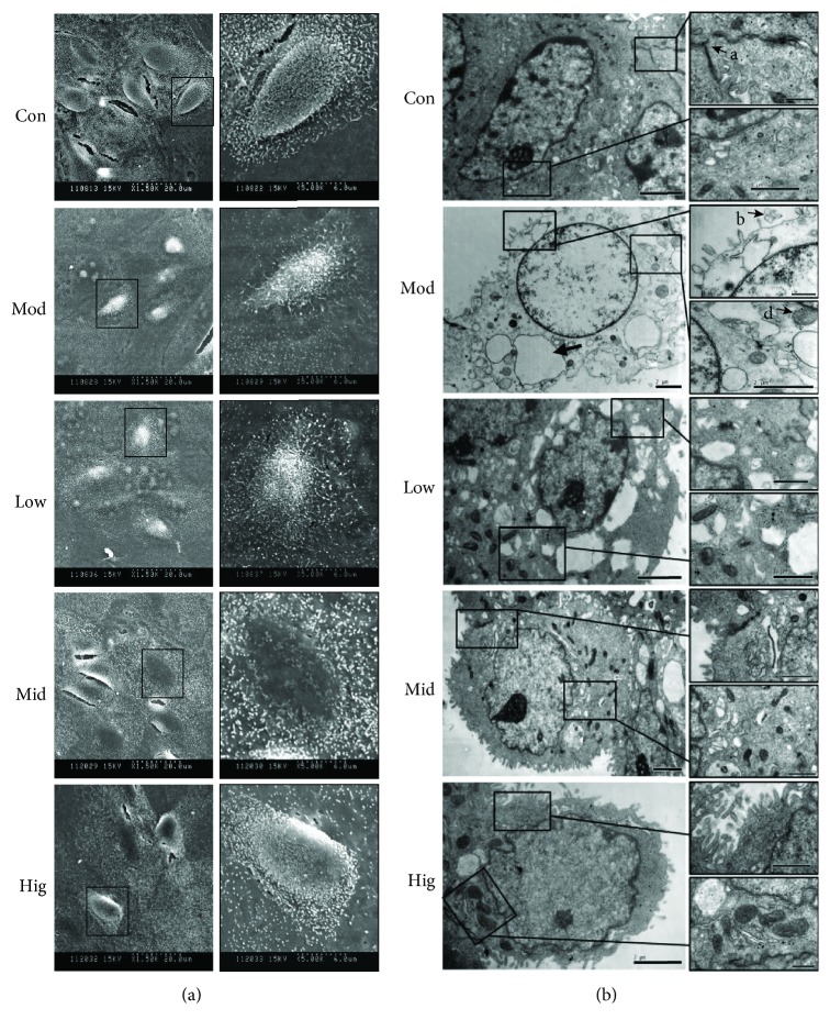
Figure 1.
CA weakened LPS-induced structural damage of bMEC. The cell microstructure of bMEC after incubation with LPS or different concentrations of CA. Cells were pretreated with indicated concentrations (10, 25, and 50 μg/mL) of CA or serum-free media for 3 h before stimulation with LPS (50 μg/mL) for 12 h; then, the microstructure was observed by SEM and TEM. (a) Compared with the Con group, the phenomenon of cell crimple and cell microvilli disorders was obviously in the Mod group; however, these changes could be improved by CA in a dose-dependent manner. The images display the view in the left panel at 1500 times actual size and the view in the right panel at 5000 times actual size. (b) The swelling or rupture of microvilli (B), extreme expansion of endoplasmic reticulum (C), and swelling and hazy of mitochondrion (D) exist in the Mod group cells, and the electron density of cytoplasmic matrix structure was more lower than that of the other groups. CA significantly improved the above features, especially the medium and high dosage. The cell tight junction in the Hig group was clearly observed as in the Con group (A), which almost disappeared in the Mod group. Con: cells without any processing; Mod: cells treated with LPS (50 μg/mL) only; Low: cells treated with LPS (50 μg/mL) after incubation with 10 μg/mL CA; Mid: cells treated with LPS (50 μg/mL) after incubation with 25 μg/mL CA; Hig: cells treated with LPS (50 μg/mL) after incubation with 50 μg/mL CA.

