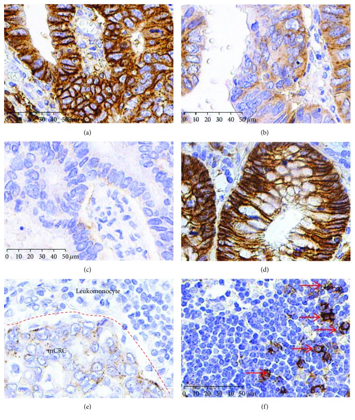
Figure 2.
SDC1 decreased both in CRCs and metastatic lymph nodes (mCRC). Immunohistochemical staining for SDC1 in CRCs. (a) High expression, (b) moderate expression, (c) low expression, (d) adjacent non-neoplastic colonic epithelium (high expression), and (e) metastatic lymph nodes (low expression). (f) SDC1 staining in nonmetastatic lymph node as negative control (nonstaining cells, T and B cells) and positive control (red arrow, plasmocytes). Scale bars: 50 μm.

