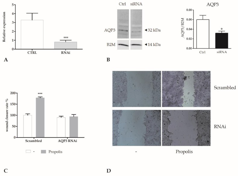
Figure 3.
The genetic silencing of AQP3 abolishes the wound closure induced by propolis. (A). Expression of AQP3 gene in HaCaT cells after RNA interference (RNAi). The mRNA quantity of AQP3 was calculated by qRT-PCR and is expressed as mean relative expression ± SD (n = 3, * p < 0.001, t-test). (B) AQP3 protein expression in HaCaT cells (CTRL) after AQP3 RNAi (RNAi). Blots representative of three were shown. Lanes were loaded with 30 μg of proteins, then probed with anti-AQP3 rabbit polyclonal antibody and managed as described in the Materials and Methods. The same blots were stripped and re-probed with anti-beta-2-microglobulin (B2M) antibody, as housekeeping (* p < 0.001, t-test). (C) Measurements of wound closure in scrambled cells or in cells exposed to RNAi for AQP3 (AQP3 RNAi), in the presence or not of 0.001% propolis, calculated as the difference between wound width at 0 and 24 h. Bars show mean ± SD of two independent experiments, each with n = 25. The mean of the control was set to 100 (*** p < 0.001, Bonferroni post-test). (D) Micrographs of scratch wounded HaCaT monolayers. Scrambled cells or cells exposed to RNAi for AQP3 (AQP3 RNAi) were incubated in the presence of 0.001% propolis and then stained with blue toluidine and observed 24 h after wounding.

