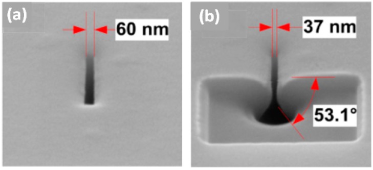
Figure 10.
SEM micrographs of a nanopore (a) before and (b) after thermal oxidation-induced shrinkage. FIB cutting was conducted to view the cross-sectional morphology of the shrunk pyramidal nanopore (reprinted with permission from [108], copyright 2013, IOP Publishing).

