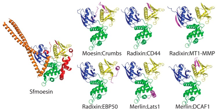
Figure 7.
Montage of merlin-ERM proteins interacting with peptides mimicking binding partner proteins. Left panel shows the Sfmoesin crystal structure [55] as a reference with domains colored as per Figure 1. On the right, individual panels show FERM:peptide complexes with the FERM colored as per Figure 1 and the peptides shown in magenta. The complexes are: moesin:Crumbs (PDB accession 4YL8 [107]); radixin:CD44 (2ZPY [104]); radixin:MT1-MMP (3X23 [106]); radixin:EBP50 (2D10 [108]); merlin:Lats1 (4ZRK [62]).; and merlin:DCAF1 (4P7I [111]). We note that for all complexes, with the exception of the radixin:MT1-MMP complex, the structure of the bound peptide mimics a portion of the CTD domain in the Sfmoesin structure (compare individual complex panels with the Sfmoesin structure). All structures are oriented the same way for direct comparison. The panels were rendered in PyMOL [63].

