Key Points
Question
Can a prognostic model predict swallowing recovery and inform the need for enteral tube feeding after dysphagic ischemic stroke?
Findings
In this single-center derivation and multicenter validation study of 279 adults with severe dysphagia, a scoring system based on 5 factors (age, stroke severity on admission, stroke location, initial risk of aspiration, and initial impairment of oral intake) predicted the recovery of functional oral intake 7 (an indication for nasogastric tube feeding) and 30 days (an indication for percutaneous endoscopic gastronomy feeding) after stroke.
Meaning
The Predictive Swallowing Score, available as the “PRESS calc” smartphone application, provided accurate predictions and could be used to guide decisions about enteral tube feeding.
Abstract
Importance
Predicting the duration of poststroke dysphagia is important to guide therapeutic decisions. Guidelines recommend nasogastric tube (NGT) feeding if swallowing impairment persists for 7 days or longer and percutaneous endoscopic gastrostomy (PEG) placement if dysphagia does not recover within 30 days, but, to our knowledge, a systematic prediction method does not exist.
Objective
To develop and validate a prognostic model predicting swallowing recovery and the need for enteral tube feeding.
Design, Setting, and Participants
We enrolled participants with consecutive admissions for acute ischemic stroke and initially severe dysphagia in a prospective single-center derivation (2011-2014) and a multicenter validation (July 2015-March 2018) cohort study in 5 tertiary stroke referral centers in Switzerland.
Exposures
Severely impaired oral intake at admission (Functional Oral Intake Scale score <5).
Main Outcomes and Measures
Recovery of oral intake (primary end point, Functional Oral Intake Scale ≥5) or return to prestroke diet (secondary end point) measured 7 (indication for NGT feeding) and 30 (indication for PEG feeding) days after stroke.
Results
In total, 279 participants (131 women [47.0%]; median age, 77 years [interquartile range, 67-84 years]) were enrolled (153 [54.8%] in the derivation study; 126 [45.2%] in the validation cohort). Overall, 64% (95% CI, 59-71) participants failed to recover functional oral intake within 7 days and 30% (95% CI, 24-37) within 30 days. Prolonged swallowing recovery was independently associated with poor outcomes after stroke. The final prognostic model, the Predictive Swallowing Score, included 5 variables: age, stroke severity on admission, lesion location, initial risk of aspiration, and initial impairment of oral intake. Predictive Swallowing Score prediction estimates ranged from 5% (score, 0) to 96% (score, 10) for a persistent impairment of oral intake on day 7 and from 2% to 62% on day 30. Model performance in the validation cohort showed a discrimination (C statistic) of 0.84 (95% CI, 0.76-0.91; P < .001) for predicting the recovery of oral intake on day 7 and 0.77 (95% CI, 0.67-0.87; P < .001) on day 30, and a discrimination for a return to prestroke diet of 0.94 (day 7; 95% CI, 0.87-1.00; P < .001) and 0.71 (day 30; 95% CI, 0.61-0.82; P < .001). Calibration plots showed high agreement between the predicted and observed outcomes.
Conclusions and Relevance
The Predictive Swallowing Score, available as a smartphone application, is an easily applied prognostic instrument that reliably predicts swallowing recovery. It will support decision making for NGT or PEG insertion after ischemic stroke and is a step toward personalized medicine.
This validation study describes the development of a prognostic model to predict swallowing recovery and guide the decision for enteral tube feeding in people with dysphagia after ischemic stroke.
Introduction
Swallowing difficulties affect around half of the estimated worldwide 3 to 6 million people who have a stroke annually.1,2 Dysphagia can impair safe oral intake, leading to malnutrition, dehydration, aspiration pneumonia, and poor poststroke outcomes.3 To prevent these complications, clinicians need to decide within the first 48 hours whether enteral tube feeding should be established.4 However, nasogastric tube (NGT) and percutaneous endoscopic gastrostomy (PEG) feeding are not without risk and not all individuals with a stroke would benefit from these procedures. Nasogastric tubes can be associated with tube misplacement, local ulcerations, discomfort, and the need to restrain.5,6 Percutaneous endoscopic gastronomies have an up to 10% risk of major complications, including infection, bleeding, and perforation.5
Guidelines recommend enteral tube feeding with NGTs if oral intake is not likely to recover within 7 days, whereas PEG is preferred if swallowing disturbances are likely to persist for more than 30 days.6,7,8 Therefore, clinicians need to predict the duration of impaired oral intake to make the right decision. There is high variability in decision making for using feeding tubes because of the lack of a systematic prediction method.9 Despite the practical need for a prognostic instrument,9 swallowing recovery prediction remains imprecise, mainly relying on a physician’s subjective experience and risk assessment.
Previous studies identified various risk factors for prolonged swallowing problems, including the National Institutes of Health Stroke Scale (NIHSS),10,11,12,13,14 bilateral infarcts,10,11,15 signs of aspiration,11,15,16 and age.13,15 We have recently demonstrated that infarctions of the frontal operculum and insular cortex are associated with impaired recovery of dysphagia.14,17 To our knowledge, an externally validated instrument to synthesize these single variables into an individualized prediction of swallowing recovery is not available.
We describe the development and validation of the Predictive Swallowing Score (PRESS), an easily applicable prognostic model for predicting the recovery of functional swallowing after ischemic stroke. From a clinical perspective, we aimed to predict recovery after 7 and 30 days to guide the decisions for early enteral tube feeding with NGT and PEG placement.
Methods
Study Population
We developed the prognostic model in a single-center prospective observational cohort study (derivation cohort) of people with ischemic stroke who were admitted to the comprehensive stroke center in St Gallen, Switzerland, between January 2011 and December 2014. We validated the model in a separate multicenter prospective observational cohort study (validation cohort) at 5 Swiss stroke centers (St Gallen, Aarau, Basel, Bern, and Lugano), between July 2015 and March 2018. The validation data were split into internal (St Gallen) and external (all other centers) categories.
To investigate people who might be later considered for enteral tube feeding, we only included individuals with ischemic stroke and a severe impairment of oral intake (Functional Oral Intake Scale [FOIS] score <5, described later) at the initial swallowing evaluation. Participants with a late admission (>48 hours after stroke), late initial swallowing assessment (>4 days after stroke), preexistent dysphagia, or stroke recurrence during the study were excluded. The local ethical committees approved the derivation and validation study protocols and participants gave written informed consent. For participants who lacked the capacity to consent, we identified a consultee and obtained written informed assent from them.
Procedures
Study flowcharts are displayed in eAppendix 1 in the Supplement. In the derivation cohort, swallowing evaluations were performed within a median of 1 day (interquartile range [IQR], 1-2 days) after a stroke, after more than 1 week (median, 8 days; IQR, 7-9 days), at discharge (median, 12 days; IQR, 6-17 days), and after more than 30 days (median, 37 days; IQR, 33-43 days) as described previously.17 In between these prespecified assessments, oral intake was evaluated during regular daily speech and language therapist (SLT) visits. Neurological data were collected at admission and at discharge. In the multicenter validation study, participants received neurological and swallowing evaluations at baseline within 4 days of stroke onset (median, 1 day; IQR, 1-2 days), after 7 days (median, 7 days; IQR, 6-8 days), and after 30 days (median, 30 days; IQR, 29-34 days).
Speech and language therapists performed comprehensive initial and follow-up swallowing assessments using a standardized procedure as reported previously17 and described in eAppendix 2 in the Supplement. All other assessments were done by neurologists. To calculate PRESS, a neurologist determined age, stroke severity, and infarct location and SLTs assessed the initial risk of aspiration and impairment of oral intake.
Definitions
We used FOIS, a reliable and validated outcome parameter, to measure adequate oral intake.18 The FOIS ranges from level 1 (nothing by mouth) to level 7 (a full unrestricted oral diet). The evaluation was done by SLTs and was based on the level of oral intake or food and liquid consistency recommended by an objective swallow assessment. Severe impairment of oral intake was defined as an oral diet of a single consistency or worse (FOIS score <5) because previous studies suggested that energy and protein intake was reduced by 40%19 and gross fluid intake was insufficient20,21 in these individuals and they would benefit from enteral tube feeding if swallowing problems persisted for at least 7 days.
The primary end point was the persistence of severely impaired oral intake (FOIS score <5) at follow-up examinations based on guidelines that recommended starting enteral tube feeding if oral intake is severely impaired for 7 days (indication for NGT feeding) and the preference for PEG feeding if oral intake is expected to remain impaired for 30 days (indication for PEG feeding) after stroke.6,7,8 Participants were considered to be recovered if FOIS score was more than 4 at an evaluation. The secondary end point was a return to the prestroke diet.
The risk of aspiration was evaluated by SLTs with the Any 2 scale22 and the 50-mL water swallow test,23 2 easily applicable methods that can be assessed at bedside. Stroke severity was measured by neurologists with the NIHSS. Stroke size (ASPECTS method)24 and dependency (modified Rankin Scale [mRS]) were evaluated by neurologists and analyzed as numerical variables without a prespecified cutoff. The severity of aphasia, dysarthria, facial weakness, and loss of consciousness was evaluated and scored (ranging from 0 = not present to 2 or 3 = severe) according to the NIHSS.
Development of the Statistical Model
We searched the literature for predictors of dysphagia recovery that can be easily assessed in different settings with various clinical expertise, are usually obtained within the first 48 hours after admission, and are part of the basic workup of people with stroke and dysphagia. We identified 5 commonly reported predictors: NIHSS score at admission,10,11,12,13,14 bilateral infarcts,10,11,15 signs of aspiration,11,15,16 age,13,15 and lesion location.14,17 We additionally performed a univariable analysis with Cox proportional hazards regression within the derivation cohort to identify factors that are associated with dysphagia recovery and, to our knowledge, have not been reported in previous studies.
For model development, we chose 11 outcome predictors that have either been consistently (≥2 studies) identified in previous research or were significant (P < .05) in the univariable analysis. First, all candidate variables were included in the Cox proportional hazards regression analysis (eAppendix 3 in the Supplement). Only 1 model is needed to predict 7-day and 30-day risk because Cox proportional hazards regression assumes that the risk of each event is proportional through time.25 Next, the full model was simplified with a backward selection procedure based on the Akaike information criterion or lack of statistical significance. The Akaike information criterion estimates the quality of each statistical model and provides a means of model selection by reducing overparameterization.26 Follow-up was censored at recovery of functional oral intake (FOIS score >4), death, the last follow-up assessment, and after day 40.
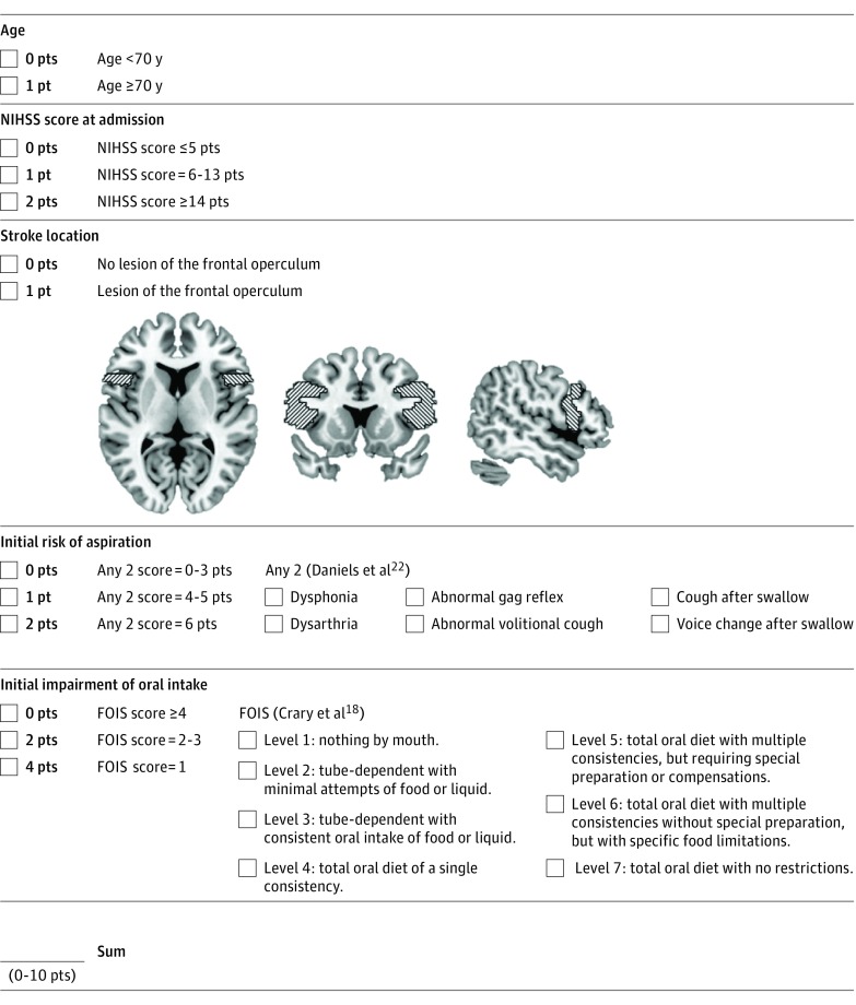
Lastly, ordinal variables were converted to categorical variables by determining significant cutoffs: an age older than 70 years; NIHSS score of 0 to 5, 6 to 13, or 14 points or greater; an initial Any 2 scale score of 1 to 3, 4 to 5, or 6 points; and an initial FOIS level of 4 or more, 2 to 3, or 1. These cutoffs were obtained by performing univariable regression analyses for each cutoff or their combination and choosing those with the highest significance. The final scoring system was developed by assigning points (0/1 or 0/1/2) to the factor levels, and, to simplify calculations, weighting them with their β-coefficients multiplied by −2 and rounding to the nearest integer. Data are presented either as number and percentage for categorical variables or as median and IQRs for ordinal and continuous variables.
Validation of the Statistical Model
The predictive accuracy of the model was determined prospectively in the multicenter validation cohort with discrimination and calibration. Model discrimination (ie, the degree to which a model differentiates between individuals with positive and negative outcomes) was calculated with the C statistic. Calibration (ie, the agreement between predicted and observed risks) was assessed with calibration plots for day 7 and day 30 and with the Hosmer-Lemeshow test.
A prespecified subgroup analysis was done in people with brainstem stroke. Predictive accuracy was compared with a previously described model for PEG placement.13 Analyses were done with R, version 3.2.3 (R Foundation). This study is reported in compliance with TRIPOD standard guidelines for prediction models.27
Results
We included 153 people in the derivation cohort and 126 in the validation cohort (64 internal, 62 external validation). Clinical characteristics are summarized in Table 1. Participants had no or minimal prestroke disability (median prestroke mRS, 0; IQR, 0-1) but were severely affected by the stroke (pooled cohort: median NIHSS score at admission, 13; IQR, 7-18). All had severe impairment of oral intake (FOIS score <5; median FOIS, 2; IQR, 1-4) and a severe risk of aspiration (median Any 2 score, 4; IQR, 3-6) at the initial swallowing examination.
Table 1. Clinical Characteristics.
| Variable | Cohort, No. (%) | ||
|---|---|---|---|
| Derivation (n = 153) |
Internal Validation (n = 64) | External Validation (n = 62) | |
| Sex | |||
| Male | 74 (48) | 37 (58) | 37 (60) |
| Female | 79 (52) | 27 (42) | 25 (40) |
| Age, y, median (IQR) | 79 (67-85) | 76 (65-85) | 74 (68-83) |
| Dependency (mRS) before admission, median (IQR) | 0 (0-1) | 0 (0-1) | 0 (0-1) |
| Stroke severity (NIHSS) at admission, median (IQR) | 12 (6-18) | 16 (11-19) | 10 (7-15) |
| Stroke laterality | |||
| Left | 72 (47) | 30 (47) | 34 (55) |
| Right | 62 (41) | 28 (44) | 21 (34) |
| Bilateral | 19 (12) | 6 (9) | 7 (11) |
| Stroke location | |||
| Cortical | 123 (80) | 51 (80) | 41 (66) |
| Subcortical | 118 (77) | 51 (80) | 43 (69) |
| Cerebellar | 16 (10) | 1 (2) | 4 (7) |
| Brainstem | 17 (11) | 6 (9) | 14 (23) |
| Affected arterial territory | |||
| Cerebral artery | |||
| Middle | 129 (84) | 57 (89) | 46 (74) |
| Anterior cerebral | 14 (9) | 7 (11) | 3 (5) |
| Posterior cerebral | 18 (12) | 5 (8) | 6 (10) |
| Basilar | 12 (8) | 3 (5) | 8 (13) |
| Vertebral | 6 (4) | 3 (5) | 11 (18) |
| Thrombolysis | 75 (49) | 40 (63) | 33 (53) |
| Stroke etiology | |||
| Small-vessel occlusion | 11 (7) | 4 (6) | 5 (8) |
| Large-artery atherosclerosis | 33 (22) | 11 (17) | 14 (23) |
| Cardioembolism | 78 (51) | 31 (48) | 20 (32) |
| Other determined origin | 5 (3) | 8 (13) | 2 (3) |
| Undetermined etiology | 26 (17) | 10 (16) | 21 (34) |
| Initial swallowing evaluation, median (IQR)a | |||
| FOIS score | 2 (1-4) | 2 (1-4) | 1 (1-4) |
| Positive 50-mL water swallow test, No. (%) | 98 (64) | 64 (100) | 60 (97) |
| Any 2 test score | 4 (3-6) | 4 (4-6) | 5 (4-6) |
| GUSS score | Not performed | 3 (2-4) | 5 (2-9) |
| PHAD score | Not performed | 62 (39-70) | 67 (56-72) |
| PRESS score | 5 (3-8) | 6 (4-9) | 6 (4-8) |
| PEG score | 2 (1-2) | 2 (2-3) | 2 (1-2) |
| Stroke outcome at 30 d or at dischargeb | |||
| Dependency (mRS), median (IQR) | 4 (2-5) | 4 (3-5) | 4 (3-4) |
| Institutionalization | 145 (95) | 45 (70) | 45 (73) |
| Pneumonia | 20 (13) | 24 (38) | 16 (26) |
| Death | 8 (5) | 11 (17) | 6 (10) |
Abbreviations: FOIS, Functional Oral Intake Scale; GUSS, Gugging Swallowing Screen; IQR, interquartile range; mRS, modified Rankin scale; NIHSS, National Institutes of Health Stroke Scale; PEG, percutaneous endoscopic gastronomy; PHAD, Parramatta Hospitals' Assessment of Dysphagia, PRESS, Predictive Swallowing Score.
Swallowing evaluations and outcome assessments at day 30 were performed by speech language therapists, and all other evaluations were done by neurologists.
The stroke outcome was determined at a 30-day follow-up visit in the validation cohorts and at discharge in the derivation cohort. The number of missing values per parameter is given in eAppendix 4 in the Supplement.
A follow-up visit after 7 days was completed in 120 people (95%) (4 [3.2%] died and 2 [1.5%] were lost to follow-up) and after 30 days in 107 people (85%) (11 [8.7%] died and 8 [6.3%] were lost to follow-up) in the validation cohort. Data were available for all outcome parameters and for 99% of the clinical variables (eAppendix 4 in the Supplement).
Clinical Course of Swallowing Recovery
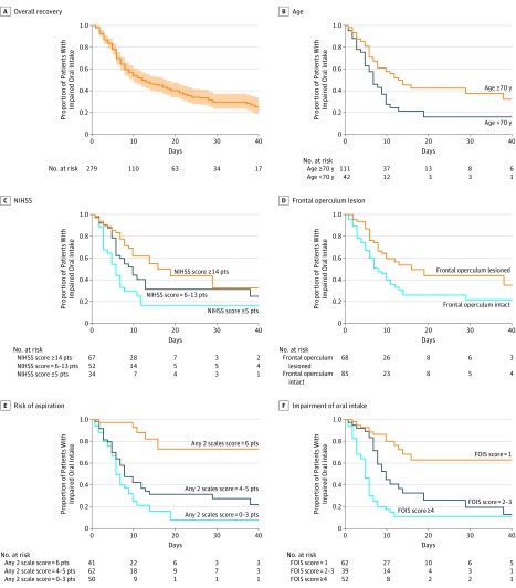
The median time to recovery of oral intake in the combined cohort (n = 279) was 12 days (95% CI, 8-16 days; Figure 1A). After 7 days, 64% (95% CI, 59-71) of participants with an initial impairment had persistent swallowing impairment and would have qualified to receive NGT feeding, according to the guidelines. After 30 days, oral intake continued to be insufficient for 30% of participants (95% CI, 24-37) and they would have qualified for PEG placement. Ninety-four percent of participants (95% CI, 88-97) failed to return to their prestroke diet after 7 days and 66% (95% CI, 57-75) after 30 days.
Figure 1. Kaplan-Meier Estimates of Time to Recovery of Oral Intake.
A, Plot of the overall clinical course of swallowing recovery (95% confidence intervals are shaded in red). B-F, Kaplan-Meier estimates for individual predictors in the Predictive Swallowing Score (PRESS) model. Separate lines are displayed for each cutoff used in PRESS. FOIS indicates Functional Oral Intake Scale; NIHSS, National Institutes of Health Stroke Scale.
A longer time to recovery of oral intake was independently associated with poor outcomes after stroke (ie, aspiration pneumonia [adjusted hazards ratio (aHR), 0.42, 95% CI, 0.27-0.66; P < .001], increased mortality [aHR, 0.06; 95% CI, 0.01-0.41; P = .01], dependency [mRS] at discharge or at the 30-day follow-up [aHR, 0.67 per point; 95% CI, 0.60-0.75; P < .001], and institutionalization [aHR, 0.52; 95% CI, 0.33-0.81; P = .004]). These results were obtained after statistical correction for sex, age, prestroke dependency, stroke severity, and stroke etiology using Cox proportional hazards models.
Model Development
The univariable analysis in the derivation cohort (n = 153) identified 6 novel predictors in addition to those 5 that were consistently reported in previous literature (eAppendix 5 in the Supplement). Five predictors remained in the final simplified model: initial impairment of oral intake (measured with FOIS), lesion of the frontal operculum, initial risk of aspiration (measured with the Any 2 test), age, and NIHSS score at admission (Figure 1). Assigning point values to these items, an integer-based calculation system was developed and termed PRESS (Figure 218,22), ranging from 0 to 10 points.
Figure 2. Calculation of the Predictive Swallowing Score.
FOIS indicates Functional Oral Intake Scale; NIHSS, National Institutes of Health Stroke Scale.
Model Performance
PRESS was a significant predictor of swallowing recovery (hazard ratio [HR], 0.73 per point; 95% CI, 0.65-0.81; P < .001) in the validation cohort (n = 126). Model performance for predicting impaired oral intake on day 7 and day 30 after stroke showed a C statistic of 0.84 (95% CI, 0.76-0.91; P < .001) and 0.77 (95% CI, 0.67-0.87; P < .001), respectively. In the internal validation cohort, discrimination was 0.86 (95% CI, 0.77-0.96; P < .001) and 0.82 (95% CI, 0.69-0.94; P < .001), respectively; in the external validation cohort, discrimination was 0.83 (95% CI, 0.71-0.95; P < .001) and 0.73 (95% CI, 0.59-0.87, P = .007), respectively.
Calibration plots showed good agreement between predicted and observed outcomes (calibration slopes ranging from 0.91-1.17; eAppendix 6 in the Supplement). The Hosmer-Lemeshow test did not suggest any significant overprediction or underprediction (eAppendix 6 in the Supplement).
We performed a prespecified subgroup analysis of people with brainstem strokes (20 [15.9%]) in the validation cohort. The C statistic after 7 and 30 days for impairment of oral intake was 0.87 (95% CI, 0.71-1.00; P = .01) and 0.77 (95% CI, 0.53-1.00; P = .08), respectively. The Hosmer-Lemeshow test did not detect miscalibration.
We also compared the performance of PRESS with a previously described model of PEG placement.13 This previously reported model had a worse performance in the validation cohort on day 7 (C statistic, 0.54; 95% CI, 0.44-0.65; P = .46) and on day 30 (C statistic, 0.54; 95% CI, 0.41-0.67; P = .52). Calibration slopes ranged from −0.01 to 0.65, suggesting poor agreement between predicted and observed data (eAppendix 7 in the Supplement). A formal comparison of PRESS with this previously proposed model showed that PRESS had substantially better discrimination on day 7 (0.84 [95% CI, 0.76-0.91] vs 0.54 [95% CI, 0.44-0.65]; P < .001) and day 30 (0.77 [95% CI, 0.67-0.87] vs 0.54 [95% CI, 0.41-0.67]; P < .001).
As a secondary end point, we determined the model performance for predicting a return to a prestroke diet. In the combined validation cohort, the C statistic was 0.94 (95% CI, 0.87-1.00; P < .001) on day 7 and 0.71 (95% CI, 0.61-0.82; P < .001) on day 30. In the internal validation cohort, discrimination was 0.92 (95% CI, 0.78-1.00; P = .02) and 0.74 (95% CI, 0.59-0.88; P = .01); in the external validation cohort, discrimination was 0.95 (95% CI, 0.89-1.00; P = .003) and 0.70 (95% CI, 0.55-0.85; P = .02). Calibration plots, slopes of the calibration regression line, and nonsignificant Hosmer-Lemeshow test results suggested good calibration for predicting a return to a prestroke diet (eAppendix 6 in the Supplement).
Model Prediction
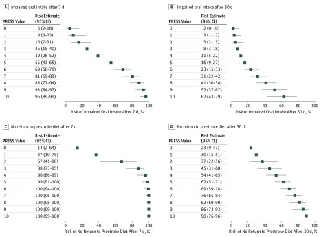
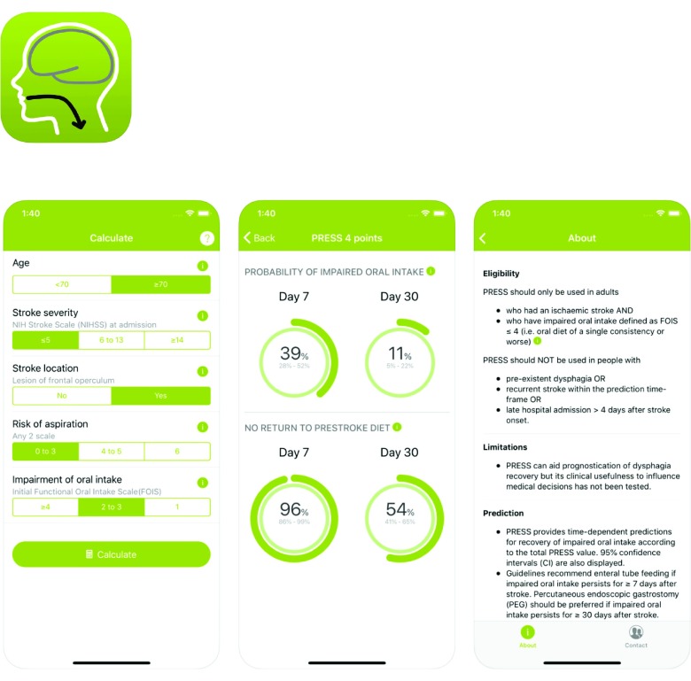
The prediction estimates for every PRESS cutoff are displayed in Figure 3. The predictions for the risk of persistent impairment of oral intake range from 5% (PRESS = 0) to 96% (PRESS = 10) on day 7 and from 2% to 62% on day 30 after stroke, covering the spectrum from rapid to prolonged swallowing recovery. An example calculation is demonstrated in eAppendix 8 in the Supplement. The sensitivity, specificity, and positive and negative predictive values of different PRESS cutoffs for predicting the impairment of oral intake or a return to a prestroke diet are described in eAppendix 9 in the Supplement. We provide a free smartphone app to facilitate PRESS calculation at bedside (Figure 4).
Figure 3. Prediction Estimates of Swallowing Recovery According to Predictive Swallowing Score (PRESS) Values.
A and B, Risk estimates for impaired oral intake 7 days (indication for nasogastric tube feeding) and 30 days (indication for percutaneous endoscopic gastronomy feeding) after stroke. C and D, Risk estimates for a failed return to a prestroke diet. Horizontal lines are 95% confidence intervals.
Figure 4. Predictive Swallowing Score (PRESS) Smartphone Graphic App.
A free smartphone and tablet app called “PRESS calc” is available to facilitate bedside examinations and prediction. It is available on Apple iOS (http://itunes.apple.com/us/app/press-calc/id1401176212) and Google Android (https://play.google.com/store/apps/details?id=ch.kssg.press).
Discussion
We developed and validated a prognostic model to predict swallowing recovery and to guide the decision for enteral tube feeding in people with dysphagia after ischemic stroke. Such a tool can have immediate practical implications for treating physicians as PRESS can be easily estimated at bedside. The model validation established good discrimination and calibration for predicting the recovery of functional oral intake and a return to a prestroke diet.
We also characterized the clinical course of swallowing within the first month after stroke. In previous studies, the incidence of dysphagia after stroke ranged widely from 2% to 33% after 1 month28,29 and from 0.4% to 50% after 6 months.28,29,30 Contrary to previous reports that focused on long-term swallowing outcomes,28,29,30 we characterized swallowing dynamics within the first weeks after stroke. Our results show that almost two-thirds of people with an initially severe dysphagic stroke do not recover functional oral intake within 7 days and therefore would benefit from NGT feeding. Nearly one-third does not recover within 30 days and would qualify for PEG feeding. Almost no individuals with a severe dysphagic stroke return to their prestroke diet within a week and two-thirds continue to require diet modification 1 month after stroke. This will be helpful in advising people with stroke and their relatives and might offer realistic expectations for the provision of health care services.
We were also able to show that prolonged recovery of functional swallowing is independently associated with poor outcomes after stroke, even after correction for stroke severity, etiology, and other clinical parameters. This result highlights the importance of developing strategies to accelerate neuronal repair mechanisms of dysphagia (eg, high-intensity speech-language therapy31 or noninvasive brain stimulation32). More efforts are needed to prevent malnutrition and dehydration in those with prolonged dysphagia, as this could be responsible for poor clinical outcomes.33
Strengths and Limitations
This study has several strengths. We assessed a comprehensive set of predictors in a large prospective cohort of people with severely dysphagic stroke. The factors that are included in PRESS are plausible; older age34 and specific infarct location17,35 might disrupt perilesional plasticity, more severe neurological deficits can interfere with the multisensory process of swallowing recovery, and the severity and type of initial swallowing impairment are likely to influence dysphagia recovery (eAppendix 10 in the Supplement). Successful prospective internal and external validation demonstrated the generalizability of the model in a Central European population despite small baseline differences between the cohorts. The validation cohort was adequately powered to show that PRESS had good discrimination. This is indicated by the 95% confidence intervals of concordance statistics, which exceeded 0.60 for both end points in the validation cohort. PRESS performed markedly better than a previously proposed model for PEG placement13 and this improvement is most likely due to integrating swallowing parameters and specific infarct location into the model.
There are some limitations. First, the results are only applicable to ischemic stroke, as we would expect different trajectories of recovery after hemorrhagic stroke.13 Second, several participants died or were lost to follow-up, and this could cause attrition bias. However, the validation cohorts had a lower loss to follow-up compared with previous studies of people with severe stroke, potentially because of performing the 30-day follow-up in a centralized manner.36 Third, it was logistically not possible to perform face-to-face follow-up after 30 days. We used a standardized central interview by a masked rater and, when possible, obtained data directly from health workers in local hospitals and rehabilitation units (85% of patients were institutionalized). Fourthly, different imaging modalities (magnetic resonance imaging in 56%, computed tomography in 44%) were used to determine stroke location and size. This might have increased data variability and reduced the reliability to detect lesions of the frontal operculum in those with computed tomography scan results, but such an approach mimics a real-life situation, making the model applicable in clinical practice. Lastly, at times there was deviation from the perfect slope in calibration plots (eAppendix 6 in the Supplement). These deviations were limited in scope and within the estimated 95% confidence intervals.
Developing a prognostic model involves making compromises and we chose only to include predictors that were well-defined, easily measured, and routinely available. Calculating PRESS does not require using specific instrumental swallowing testing (eg, videofluoroscopic or fiberoptic evaluation) and this makes the model applicable in different countries and settings with various clinical expertise. Future research might refine predictions by including such instrumental biomarkers and advanced structural or functional neuroimaging.
Conclusions
Our results show that a simple algorithm can predict swallowing recovery and might support decision making in people with dysphagic stroke. Such an algorithm requires cross-specialty collaboration between neurologists and SLTs to determine accurate predictions. PRESS has the potential to individualize clinical care by identifying those who would benefit from enteral tube feeding and is a step toward personalized medicine.
eAppendix 1. Study flow charts.
eAppendix 2. Swallowing assessments.
eAppendix 3. Missing data.
eAppendix 4. Univariable analysis in derivation cohort.
eAppendix 5. Calibration plots for PRESS.
eAppendix 6. Performance of a model previously proposed in literature.
eAppendix 7. Example PRESS calculation.
eAppendix 8. Classification parameters for PRESS cutoffs.
eAppendix 9. Plausibility of factors included in the PRESS model.
References
- 1.Seshadri S, Wolf PA. Lifetime risk of stroke and dementia: current concepts, and estimates from the Framingham Study. Lancet Neurol. 2007;6(12):1106-1114. doi: 10.1016/S1474-4422(07)70291-0 [DOI] [PubMed] [Google Scholar]
- 2.Martino R, Foley N, Bhogal S, Diamant N, Speechley M, Teasell R. Dysphagia after stroke: incidence, diagnosis, and pulmonary complications. Stroke. 2005;36(12):2756-2763. doi: 10.1161/01.STR.0000190056.76543.eb [DOI] [PubMed] [Google Scholar]
- 3.Dávalos A, Ricart W, Gonzalez-Huix F, et al. . Effect of malnutrition after acute stroke on clinical outcome. Stroke. 1996;27(6):1028-1032. doi: 10.1161/01.STR.27.6.1028 [DOI] [PubMed] [Google Scholar]
- 4.Dennis MS, Lewis SC, Warlow C; FOOD Trial Collaboration . Effect of timing and method of enteral tube feeding for dysphagic stroke patients (FOOD): a multicentre randomised controlled trial. Lancet. 2005;365(9461):764-772. doi: 10.1016/S0140-6736(05)70999-5 [DOI] [PubMed] [Google Scholar]
- 5.Dennis M, Lewis S, Cranswick G, Forbes J; FOOD Trial Collaboration . FOOD: a multicentre randomised trial evaluating feeding policies in patients admitted to hospital with a recent stroke. Health Technol Assess. 2006;10(2):iii-iv, ix-x, 1-120. doi: 10.3310/hta10020 [DOI] [PubMed] [Google Scholar]
- 6.Kirby DF, Delegge MH, Fleming CR. American Gastroenterological Association technical review on tube feeding for enteral nutrition. Gastroenterology. 1995;108(4):1282-1301. doi: 10.1016/0016-5085(95)90231-7 [DOI] [PubMed] [Google Scholar]
- 7.Stroud M, Duncan H, Nightingale J; British Society of Gastroenterology . Guidelines for enteral feeding in adult hospital patients. Gut. 2003;52(suppl 7):vii1-vii12. doi: 10.1136/gut.52.suppl_7.vii1 [DOI] [PMC free article] [PubMed] [Google Scholar]
- 8.Wirth R, Smoliner C, Jäger M, Warnecke T, Leischker AH, Dziewas R; DGEM Steering Committee . Guideline clinical nutrition in patients with stroke. Exp Transl Stroke Med. 2013;5(1):14. doi: 10.1186/2040-7378-5-14 [DOI] [PMC free article] [PubMed] [Google Scholar]
- 9.George BP, Kelly AG, Schneider EB, Holloway RG. Current practices in feeding tube placement for US acute ischemic stroke inpatients. Neurology. 2014;83(10):874-882. doi: 10.1212/WNL.0000000000000764 [DOI] [PMC free article] [PubMed] [Google Scholar]
- 10.Kumar S, Langmore S, Goddeau RP Jr, et al. . Predictors of percutaneous endoscopic gastrostomy tube placement in patients with severe dysphagia from an acute-subacute hemispheric infarction. J Stroke Cerebrovasc Dis. 2012;21(2):114-120. doi: 10.1016/j.jstrokecerebrovasdis.2010.05.010 [DOI] [PMC free article] [PubMed] [Google Scholar]
- 11.Kumar S, Doughty C, Doros G, et al. . Recovery of swallowing after dysphagic stroke: an analysis of prognostic factors. J Stroke Cerebrovasc Dis. 2014;23(1):56-62. doi: 10.1016/j.jstrokecerebrovasdis.2012.09.005 [DOI] [PubMed] [Google Scholar]
- 12.San Luis COV, Staff I, Ollenschleger MD, Fortunato GJ, McCullough LD. Percutaneous endoscopic gastrostomy tube placement in left versus right middle cerebral artery stroke: effects of laterality. NeuroRehabilitation. 2013;33(2):201-208. [DOI] [PubMed] [Google Scholar]
- 13.Dubin PH, Boehme AK, Siegler JE, et al. . New model for predicting surgical feeding tube placement in patients with an acute stroke event. Stroke. 2013;44(11):3232-3234. doi: 10.1161/STROKEAHA.113.002402 [DOI] [PMC free article] [PubMed] [Google Scholar]
- 14.Galovic M, Leisi N, Müller M, et al. . Lesion location predicts transient and extended risk of aspiration after supratentorial ischemic stroke. Stroke. 2013;44(10):2760-2767. doi: 10.1161/STROKEAHA.113.001690 [DOI] [PubMed] [Google Scholar]
- 15.Ickenstein GW, Kelly PJ, Furie KL, et al. . Predictors of feeding gastrostomy tube removal in stroke patients with dysphagia. J Stroke Cerebrovasc Dis. 2003;12(4):169-174. doi: 10.1016/S1052-3057(03)00077-6 [DOI] [PubMed] [Google Scholar]
- 16.Ickenstein GW, Höhlig C, Prosiegel M, et al. . Prediction of outcome in neurogenic oropharyngeal dysphagia within 72 hours of acute stroke. J Stroke Cerebrovasc Dis. 2012;21(7):569-576. doi: 10.1016/j.jstrokecerebrovasdis.2011.01.004 [DOI] [PubMed] [Google Scholar]
- 17.Galovic M, Leisi N, Pastore-Wapp M, et al. . Diverging lesion and connectivity patterns influence early and late swallowing recovery after hemispheric stroke. Hum Brain Mapp. 2017;38(4):2165-2176. doi: 10.1002/hbm.23511 [DOI] [PMC free article] [PubMed] [Google Scholar]
- 18.Crary MA, Mann GDC, Groher ME. Initial psychometric assessment of a functional oral intake scale for dysphagia in stroke patients. Arch Phys Med Rehabil. 2005;86(8):1516-1520. doi: 10.1016/j.apmr.2004.11.049 [DOI] [PubMed] [Google Scholar]
- 19.Wright L, Cotter D, Hickson M, Frost G. Comparison of energy and protein intakes of older people consuming a texture modified diet with a normal hospital diet. J Hum Nutr Diet. 2005;18(3):213-219. doi: 10.1111/j.1365-277X.2005.00605.x [DOI] [PubMed] [Google Scholar]
- 20.Whelan K. Inadequate fluid intakes in dysphagic acute stroke. Clin Nutr. 2001;20(5):423-428. doi: 10.1054/clnu.2001.0467 [DOI] [PubMed] [Google Scholar]
- 21.Vivanti AP, Campbell KL, Suter MS, Hannan-Jones MT, Hulcombe JA. Contribution of thickened drinks, food and enteral and parenteral fluids to fluid intake in hospitalised patients with dysphagia. J Hum Nutr Diet. 2009;22(2):148-155. doi: 10.1111/j.1365-277X.2009.00944.x [DOI] [PubMed] [Google Scholar]
- 22.Daniels S, McAdam C, Brailey K, Foundas A. Clinical assessment of swallowing and prediction of dysphagia severity. Am J Speech Lang Pathol. 1997;6(4):17-24. doi: 10.1044/1058-0360.0604.17 [DOI] [Google Scholar]
- 23.DePippo KL, Holas MA, Reding MJ. Validation of the 3-oz water swallow test for aspiration following stroke. Arch Neurol. 1992;49(12):1259-1261. doi: 10.1001/archneur.1992.00530360057018 [DOI] [PubMed] [Google Scholar]
- 24.Barber PA, Demchuk AM, Zhang J, Buchan AM. Validity and reliability of a quantitative computed tomography score in predicting outcome of hyperacute stroke before thrombolytic therapy. ASPECTS Study Group. Alberta Stroke Programme Early CT score. Lancet. 2000;355(9216):1670-1674. doi: 10.1016/S0140-6736(00)02237-6 [DOI] [PubMed] [Google Scholar]
- 25.Vittinghoff E, Glidden DV, Shiboski SC, McCulloch CE. Regression Methods in Biostatistics. New York, NY: Springer Science & Business Media; 2012. doi: 10.1007/978-1-4614-1353-0 [DOI] [Google Scholar]
- 26.Bozdogan H. Model selection and Akaike’s Information Criterion (AIC): the general theory and its analytical extensions. Psychometrika. 1987;52(3):345-370. doi: 10.1007/BF02294361 [DOI] [Google Scholar]
- 27.Collins GS, Reitsma JB, Altman DG, Moons KGM. Transparent Reporting of a Multivariable Prediction Model for Individual Prognosis or Diagnosis (TRIPOD): the TRIPOD statement. Ann Intern Med. 2015;162(1):55-63. doi: 10.7326/M14-0697 [DOI] [PubMed] [Google Scholar]
- 28.Barer DH. The natural history and functional consequences of dysphagia after hemispheric stroke. J Neurol Neurosurg Psychiatry. 1989;52(2):236-241. doi: 10.1136/jnnp.52.2.236 [DOI] [PMC free article] [PubMed] [Google Scholar]
- 29.Smithard DG, O’Neill PA, England RE, et al. . The natural history of dysphagia following a stroke. Dysphagia. 1997;12(4):188-193. doi: 10.1007/PL00009535 [DOI] [PubMed] [Google Scholar]
- 30.Mann G, Hankey GJ, Cameron D. Swallowing function after stroke: prognosis and prognostic factors at 6 months. Stroke. 1999;30(4):744-748. doi: 10.1161/01.STR.30.4.744 [DOI] [PubMed] [Google Scholar]
- 31.Nakazora T, Maeda J, Iwamoto K, et al. . Intervention by speech therapists to promote oral intake of patients with acute stroke: a retrospective cohort study. J Stroke Cerebrovasc Dis. 2017;26(3):480-487. doi: 10.1016/j.jstrokecerebrovasdis.2016.12.007 [DOI] [PubMed] [Google Scholar]
- 32.Simons A, Hamdy S. The use of brain stimulation in dysphagia management. Dysphagia. 2017;32(2):209-215. doi: 10.1007/s00455-017-9789-z [DOI] [PMC free article] [PubMed] [Google Scholar]
- 33.Zhang J, Zhao X, Wang A, et al. . Emerging malnutrition during hospitalisation independently predicts poor 3-month outcomes after acute stroke: data from a Chinese cohort. Asia Pac J Clin Nutr. 2015;24(3):379-386. doi: 10.6133/apjcn.2015.24.3.13 [DOI] [PubMed] [Google Scholar]
- 34.Li S, Overman JJ, Katsman D, et al. . An age-related sprouting transcriptome provides molecular control of axonal sprouting after stroke. Nat Neurosci. 2010;13(12):1496-1504. doi: 10.1038/nn.2674 [DOI] [PMC free article] [PubMed] [Google Scholar]
- 35.Galovic M, Leisi N, Müller M, et al. . Neuroanatomical correlates of tube dependency and impaired oral intake after hemispheric stroke. Eur J Neurol. 2016;23(5):926-934. doi: 10.1111/ene.12964 [DOI] [PubMed] [Google Scholar]
- 36.Pendlebury ST, Chen P-J, Welch SJV, et al. ; Oxford Vascular Study . Methodological factors in determining risk of dementia after transient ischemic attack and stroke: (II) effect of attrition on follow-up. Stroke. 2015;46(6):1494-1500. doi: 10.1161/STROKEAHA.115.009065 [DOI] [PMC free article] [PubMed] [Google Scholar]
Associated Data
This section collects any data citations, data availability statements, or supplementary materials included in this article.
Supplementary Materials
eAppendix 1. Study flow charts.
eAppendix 2. Swallowing assessments.
eAppendix 3. Missing data.
eAppendix 4. Univariable analysis in derivation cohort.
eAppendix 5. Calibration plots for PRESS.
eAppendix 6. Performance of a model previously proposed in literature.
eAppendix 7. Example PRESS calculation.
eAppendix 8. Classification parameters for PRESS cutoffs.
eAppendix 9. Plausibility of factors included in the PRESS model.


