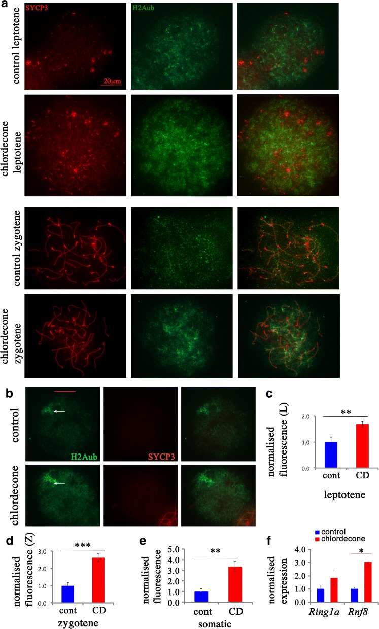
Fig. 3.
Meiosis defects are associated with altered H2Aub occupancy. a Surface spreads from E15.5 ovaries in leptotene stage from control (first row) and CD-exposed mice (second row) and in zygotene stage from control (third row) and CD-exposed (fourth row) ovaries (63X magnification). Spreads from ovaries were immunostained with anti-H2Aub (green) and anti-SYCP3 (red) antibodies. Quantitative analysis of anti-H2Aub intensity in cells in leptotene and (c) in zygotene (d) stages. b Representative images of H2Aub in somatic cells. The Barr body is shown with an arrow. Images from 4 control and 5 treatment biological replicates were used for analysis, and the normalized fluorescence intensities in somatic cells (e) are provided. The immunofluorescence was performed as described in Methods section using antibodies against H2Aub and SYCP3; the images were obtained using microscope using the fixed exposure time. The images were analyzed using ImageJ software and normalized fluorescence was calculated and the averaged value ± SEM were compared, **p < 0.01, t test, the bar represents 20 µm. The analysis was performed on at least 20 oocytes from four replicates for each group, n = 4 for each group, *p < 0.05, **p < 0.01, ***p < 0.001, t test. f The gene expression level of Rnf8 and Ring1a genes was analyzed by RT-qPCR using RNA from E15.5 ovaries from control and CD-treated animals, **p < 0.01, t test

