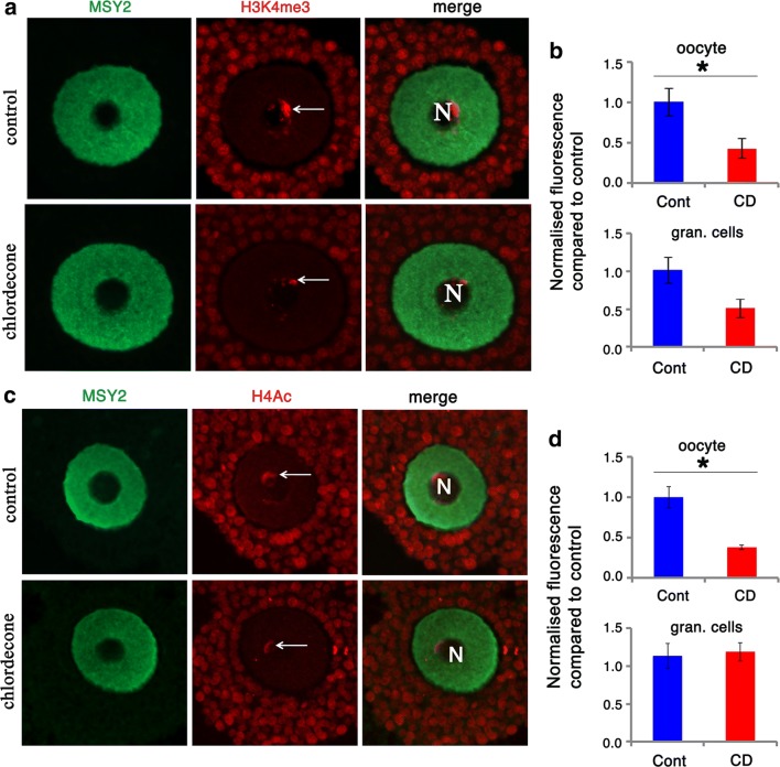
Fig. 8.
The decrease in histone H3K4me3 and H4 acetylation levels in adult ovaries following gestational CD exposure. Exposure to CD affects histone H3K4me3 and H4Ac levels in adult ovaries. a Representative images of control (top) or treated oocyte (bottom) immunostained by MSY2 (oocyte marker, green) or H3K4me3 (red) (40X magnification). b Quantitative analysis of H3K4me3 in oocytes and the surrounding granulosa cells, n = 4 for each group, *p < 0.05, **p < 0.01, t test. c Representative images of antral follicles immunostained for acetylated histone 4 (red). Note that in control samples homogenous nuclear staining is observed, while in treated samples clusters of bright dots are visible. d Quantitative analysis of H4 acetylation in granulosa cells and oocytes of control and CD-treated samples, n = 4 for each group, *p < 0.05, t test

