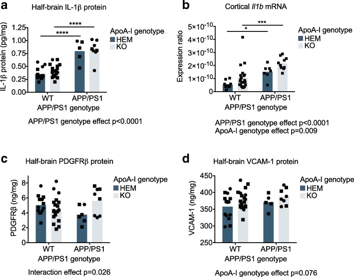
Fig. 3.
Cortical levels of pro-inflammatory protein and mRNA markers were increased in the absence of apoA-I. a IL-1β, c PDGFRβ, and d VCAM-1 protein levels were measured by ELISA in soluble half-brain homogenates; values were normalized to total protein concentration in the homogenates. b Il1b mRNA expression was measured in the cortex by qRT-PCR and normalized to β-actin expression. Points represent individual mice, and bars represent mean values. Circles represent female mice, and squares represent male mice. Omnibus analyses of apoA-I and APP/PS1 genotype effects by two-way ANOVA are displayed as exact p values below graphs. Sidak’s multiple comparisons test results are displayed within graphs as *p < 0.05, ***p < 0.001, and ****p < 0.0001. For ELISA, N = 5–19 mice per genotype were used; for mRNA, N = 7–21 mice per genotype were used. apoA-I, apolipoprotein A-I; HEM, hemizygous apoA-I genotype; KO, knockout apoA-I genotype; WT, wildtype APP/PS1 genotype; APP/PS1, transgenic APP/PS1 genotype; IL-1β, interleukin 1 beta; VCAM-1, vascular cell adhesion molecule 1; PDGFRβ, platelet-derived growth factor receptor beta

