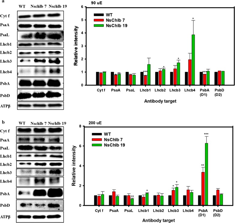
Fig. 5.
Quantitation of photosynthetic proteins via western blotting and estimation of band intensities. Cells were grown under ML (a) or HL (b), and 1.5 × 10⁸ cells were harvested on day 8 for western blotting. Band intensities for individual proteins were normalized with that of ATPβ (using the Image Lab software), and relative intensities to WT are presented on the right panel. Each data point represents the average of three independent replicates and error bars are standard errors. Asterisks indicate the significant differences between WT and each transformants (WT vs. NsChlb7, WT vs. NsChlb19) determined by Student’s t test (*P < 0.05. **P < 0.01, ***P < 0.001)

