Figure 4.

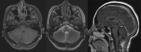
Simple and contrast magnetic resonance image, sequences weighted in T1 axial cut, T2 axial cut, and sequences contrast sagittal where no tumor residue is identified in the posterior fossa

Simple and contrast magnetic resonance image, sequences weighted in T1 axial cut, T2 axial cut, and sequences contrast sagittal where no tumor residue is identified in the posterior fossa