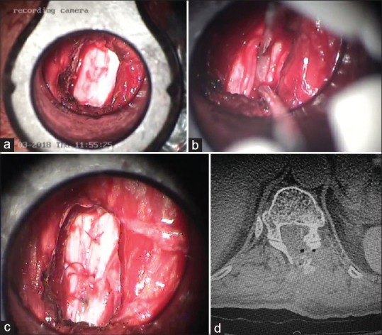
Figure 6.

(a) Visualization of the dura after hemilaminectomy using nonexpendable retractor system. (b) Tumor and roots were visualized through the tubular retractor system. (c) Dura was closed using 6-0 polyglactin interrupted sutures. (d) Postoperative computed tomography scan showing the left hemilaminectomy in minimally invasive surgery for tumor excision. Bone pieces removed during laminectomy were replaced back on the dural surface
