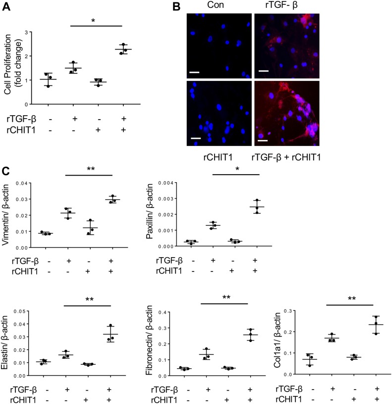
Figure 1. CHIT1 enhances TGF-β1-stimulated fibroblast responses in vitro.
NHLF were stimulated with recombinant (r) TGF-β1 (rTGF-β, 10 ng/ml) and rCHIT1 (250 ng/ml) for 24 h then the effect of rTGF-β and rCHIT1 was evaluated. (A) Fibroblast cell proliferation assay by WST-1. (B) Immunohistochemistry (IHC) using anti–α-smooth muscle actin; Con, controls with no rTGF-β or rCHIT1 stimulation. (C) Expression of markers of myofibroblasts and extracellular protein accumulation assessed by real-time quantitative reverse transcription PCR (qRT-PCR). β-actin was used as an internal control. The values in panels A and C represent mean ± SEM of triplicated evaluations in a minimum of two separate experiments. Panel B is a representative IHC in a minimum of three separate experiments. *P < 0.05, **P < 0.01, t test. Bars in panel B, 50 μM.

