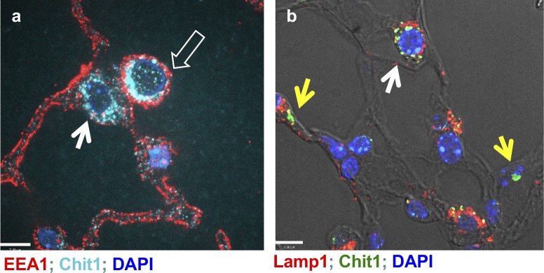
Figure S4. Intracellular localization of Chit1 expression in the bleomycin-stimulated lung.
Fluorescent IHC to localize Chit1 and early endosomal (EEA1) or lysosomal markers (Lamp1) on the lung tissue sections from bleomycin-challenged lungs. Open arrow, macrophage; closed arrow, type 2 alveolar epithelial cells; and yellow arrows, type 1 epithelial cells. (A, B) Bars in (A) and (B), 12 and 7 μm, respectively.

