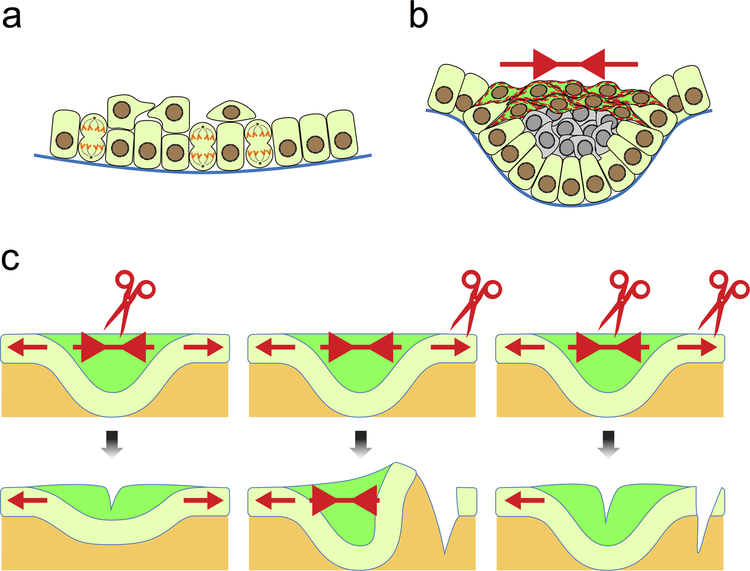
Fig. 3. Mechanical input for dental epithelial invagination.
a) Dental placode is formed as a result of vertical cell division, which generates suprabasal cells and thus the initial thickening of the epithelium. b) Basal cells (light green) at the edges of the placode intercalate with suprabasal cells, which also intercalate within themselves at the more apical portion of the placode (dark green). This creates a contractile tension (red arrows) and leads to the bending of the epithelium. The observation of thick actin bundles and strong phosphomyosin staining (shown as red dashed lines in dark green cells) reflects such tension in the epithelium. c) The tensional forces can also be detected by cutting the epithelium at different points. Cutting within the suprabasal layer relieves the contraction, resulting in a more relaxed and shallow tooth germ. Cutting through the adjacent oral epithelium causes the tooth germ to bend further as the contraction is no longer resisted. Cutting in both the suprabasal layer and the neighboring epithelium abrogates these effects, as the net force is again equivalent between the two regions.

