Figure 6.
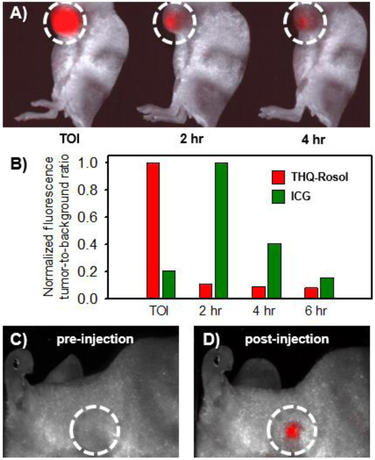
In vivo efficacy of THQ-Rosol toward lymphatic mapping applications. A) Representative images of THQ-Rosol following its administration to a GBM39 xenograft mouse model at select time points. B) The accompanying normalized fluorescence tumor-to-background ratio of THQ-Rosol (λem2 = 710 nm) compared to ICG (λem1 = 800–900 nm) at select time points (each 50 μL, 25 μM). Accumulation of THQ-Rosol in the axillary lymph node C) pre and D) post administration. TOI = time of injection. Dashed white circle circumscribes the axillary lymph node region.

