
Figure 1. Processing pipeline.
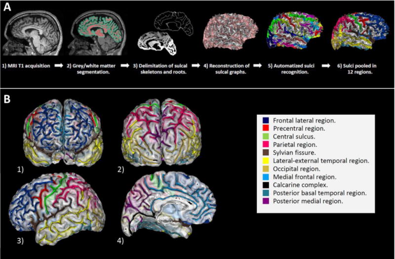
1-A : Processing steps embedded in the BrainVISA morphologist pipeline. 1-B : The twelve predetermined cortical regions investigated with local sulcation index. 1) Anterior view. 2) Posterior view. 3) Lateral view of the left hemisphere. 4) Medial view of the left hemisphere. The 12 cortical regions that have been studied are shown in color. Navy blue: Frontal dorso-lateral region. Red: Precentral region. Green: Central sulcus. Pink: Parietal region. Brown : Sylvian fissure. Yellow: Lateral – external temporal region. Gold: Occipital region. Light blue: Medial frontal region. Dark: Calcarine complex. Green blue: Posterior basal temporal region. Violet: Posterior medial region.