
Figure 5.
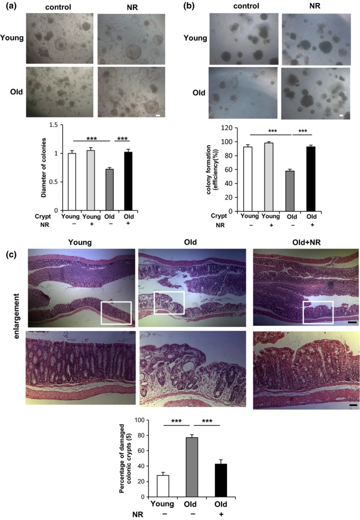
NR treatment reverses functional decline in ISCs during aging in vivo. (a) Isolated young or old colonic crypts were cultured in medium with or without 1 mM NR as indicated. The diameter of more than 70 colonies per each group was analyzed on day 5. (b) The number of colonies cultured in medium with or without 1 mM NR was assessed after 6 days of primary culture and an additional 5 days of secondary passage (4–5 wells per group). Original magnifications: ×50 (a and b). Scale bar: 100 µm (a and b). (c) Young and old mice administered with vehicle or NR (500 mg/kg) in drinking water for 6 weeks were treated by 1.5% DSS for 5 days followed by 3‐day water feeding before being sacrificed (five mice per group). The HE staining of distal colon (10 mm from middle) tissues and the percentage of damaged colonic crypts are shown. Bottom: the enlargement of the squared area. At least three different ×40 image fields per each sample were quantified. Original magnifications: ×50 and ×200 (enlargement). Scale bar: 200 and 50 µm (enlargement). Values represent the mean ± SEM. ***p < 0.001; t test. DSS: dextran sulfate sodium; ISCs: intestinal stem cells; NR: nicotinamide riboside