Figure 3.

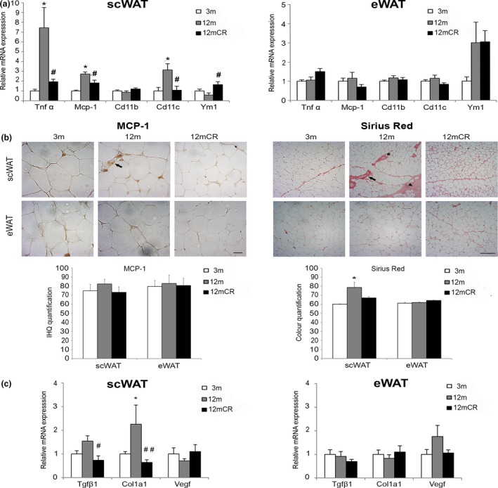
Increased fibro‐inflammation state in scWAT at early stages of aging. (a) mRNA levels of representative inflammatory genes in scWAT and eWAT in the three experimental groups. (b) Representative images of immunohistochemically analysis of MCP‐1 expression in paraffin‐embedded scWAT and eWAT from histological sections (magnification 40×, scale bar = 20μm) of the experimental groups (n = 4 animals/group). No immunoreaction was observed in the negative control treated with PBS without primary antibody (not shown). Arrows = locations of infiltrated MCP‐1‐expressing macrophages, which form crown‐like structures surrounding dead and dying adipocytes. Moreover, representative images of histological determination of fibrosis with Sirius red staining for collagen in paraffin‐embedded fat depots (magnification 10×, scale bar = 500μm) of the three experimental groups (n = 4 animals/group) are shown. Arrows = abundant fibrosis around vessels; arrowheads = collagen fibers organized in bundles containing a few adipocytes isolated from the rest of the parenchyma; asterisk = thinner collagen fibrils around adipocytes (i.e., pericellular fibrosis). (c) mRNA levels of representative fibrosis genes in scWAT and eWAT in the three experimental groups. All data are expressed as mean ± SEM (a, d: n = 7–9 animals/group). * p < 0.05, 12 m vs. 3 m; # p < 0.05, ## p < 0.01, 12mCR vs. 12 m
