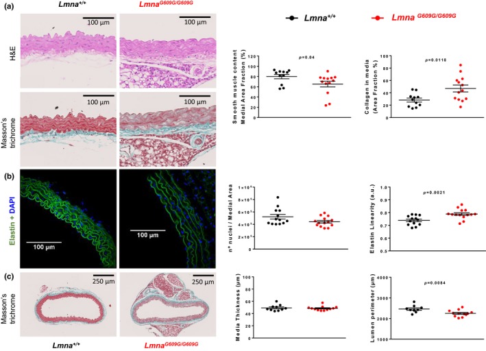
Figure 5.

Aortic media of LmnaG609G/G609G mice shows increased collagen deposition, a decreased amount of smooth muscle tissue, and altered elastin waving. (a) Histological analysis of aortic sections from LmnaG609G/G609G mice (n = 13) and Lmna+/+mice (n = 11) stained with H&E and Masson's trichrome, showing increased collagen deposition and decreased smooth muscle area in the aortic medial layer of progeroid mice. (b) Confocal microscopy images of elastin autofluorescence and DAPI nuclear staining (n = 13–14), showing increased elastin wave linearity in aortic sections from LmnaG609G/G609G mice unaccompanied by significant changes in nuclear number. (c) Morphological analysis of whole aortic sections (n = 9–12), showing no change in medial layer thickness and a decreased lumen perimeter relative to controls, indicating inward remodeling in the LmnaG609G/G609G aorta
