
Figure 1.
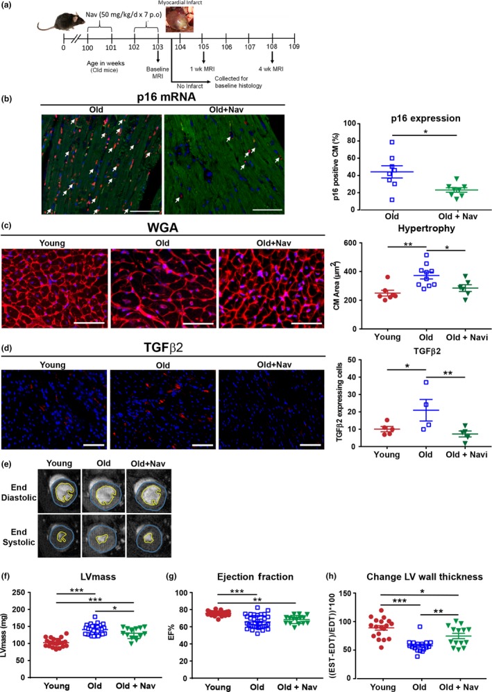
Aged mice display increased CM senescence, CM hypertrophy, increased TGFβ2 expression and functional characteristics of myocardial aging, which are attenuated by navitoclax treatment. (a) Experimental design. (b) Percentage p16+ CMs by RNA in situ hybridization. Arrow indicates p16 expressing CMs (p16 red, troponin‐C green, DAPI blue), n = 8 per group. Scale bars = 50 µm. (c) WGA staining and quantification of CM cross‐sectional area, n = 6–10 per group. (d) TGFβ2 protein expression, n = 4–5 per group. (e) Examples of individual short axis cine‐MR images. Analysis of (f) left ventricular mass and (g) ejection fraction. (h) % change in wall thickness, n = 14–34 per group. For c and d, scale bars = 100 µm. Data are mean ± SEM, ***p < 0.001; **p < 0.01; * p < 0.05 using Student's t test or one‐way ANOVA