Figure 1.

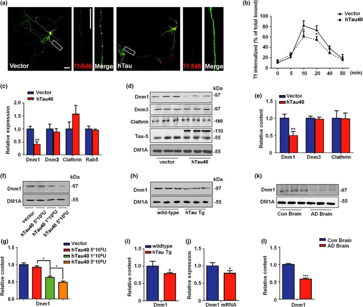
Tau interrupts synaptic endocytosis by decreasing dynamin 1. (a) Primary cortical neurons were infected with lentivirus packed hTau‐EGFP or EGFP at DIV7, and the Transferrin (Tf‐546) uptake experiments were performed at 72 hr later. The red color indicates the internalized Transferrin. Bar = 50 μm. N = 5. (b) The effects of hTau on Tf‐546 endocytosis were detected in several time points. N = 5. (c) The neurons were treated as above, and the mRNA of dynamin 1 (Dnm1), dynamin 3 (Dnm3), clathrin, and Rab5 were detected. N = 5. **p < 0.01, vs. vector. (d) The representative blots of dynamin1, dynamin3, clathrin, and Tau5 and (e) the quantification. N = 5. **p < 0.01, vs. vector. (f) The representative blots of dynamin1 in neurons that treated with different hTau lentivirus dilutions and (g) the quantification. N = 5.*p < 0.05, vs. vector. (h) The representative blots of dynamin1 in the cortex of 12 weeks hTau transgenic mice and their wild‐type and (i) the quantification. N = 6. *p < 0.05, vs. wild‐type. (j) The dynamin1 mRNA level in the cortex of 12 weeks hTau transgenic and age‐matched wild‐type mice. N = 5. *p < 0.05, vs. wild‐type. (k) The representative blots of dynamin1 in AD brain and control brain, and (l) the quantification. N = 5. *p < 0.05, vs. control
