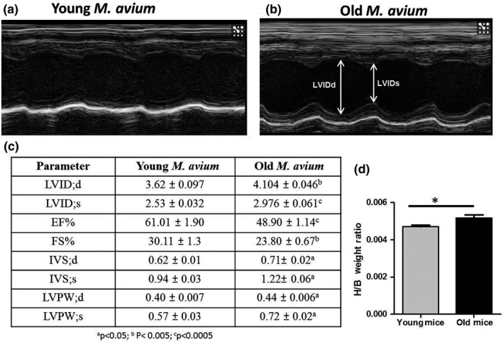
Figure 3.

Hearts of Mycobacterium avium infected old mice undergo cardiac hypertrophy. To assess heart function in vivo, 2D‐echocardiography (Vevo 2100, Visualsonics) was performed in M. avium (200 CFUs) infected young and old mice at 30 days postinfection. Representative baseline M‐mode echocardiographs from M. avium infected young (a) and old (b) mice (N = 6). (c) Atleast two M‐mode echocardiogram measurements from each mouse were used to determine the LVIDd, left ventricular end‐diastolic dimension; LVIDs, left ventricular end‐systolic dimension, ejection fraction (EF%), fractional shortening (FS%), IVSTd, interventricular septal thickness in diastole; IVSTs, interventricular septal thickness in systole; LVPWd, posterior wall thickness in diastole; LVPWDs, posterior wall thickness in systole, a p < 0.05; b p < 0.005; c p < 0.0005. (d) Graph shows the heart weight over the body weight of young and old mice (baseline), 20 mice/group (mean ± SEM; *p < 0.05)
