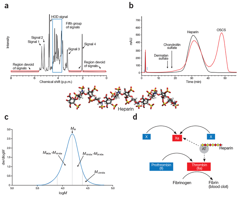
Figure 3.
Orthogonal analysis of heparin. (a) Spectroscopic analysis shows a typical 1H-NMR spectrum of heparin. The specification for heparin included the identification of five groups of signals is: Signal 1 (5.42 p.p.m.) represents H1 of GlcNAc6S/GlcNS6S; Signal 2 (5.21 p.p.m.) represents H1 of IdoA2S; Signal 3 represents H2 of GlcNS (3.28 p.p.m.); and Signal 4 represents the methyl of GlcNAc (2.05 p.p.m.). The fifth group of signals (3.75–4.55 p.p.m.), corresponding to a number of heparin protons, are surrounded by a blue box. Signals 1, 2, and 4 are of comparable intensity. Signal 3 is substantially smaller than signal 2. The intensity of the fifth group of signals are 1–3 times higher than signal 2. In addition, the spectrum profile between 3.2 p.p.m. and 4.6 p.p.m., and between 4.9 p.p.m. and 5.7 p.p.m., closely matches the reference spectrum, both in intensity and signal position. The three red boxes surround regions of the spectrum where no signals should be observed. (b) Chromatographic mobility analysis showing compiled SAX–HPLC chromatograms of heparin standard (black tracing) and the mixture of HP and OSCS (red tracing). The eluted positions of other glycosaminoglycans, like dermatan sulfate and chondroitin sulfate, are indicated by arrows. (c) Molecular weight distribution analysis, shows a typical molecular weight distribution curve of heparin. In this particular determination, the weight average molecular weight (Mw) is 15,916; the proportion of material above 24 kDa (M>24 kDa) is 8.4% and the ratio of material 8–16 kDa to 16–24 kDa (M8–16 kDa/M16–24 kDa) is 1.46. dw/dlogM (y axis) represents the fraction of the material by weight. (d) Potency analysis of a partial blood coagulation cascade, demonstrating the mechanism of action of heparin. Heparin achieves its anticoagulant activity by binding to antithrombin (AT), and the heparin/AT complex inhibits the activities of factor Xa and factor IIa to prevent the formation of fibrin from fibrinogen, which is a key step in generating blood clots. The dashed arrows depict the action sites in the blood coagulation cascade by the complex of AT/heparin.

