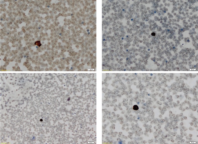
Figure 1.
Representative images of microscope slides showing CTC negativity (A and B) and CTC positivity (C and D), as represented by presence or absence of immunostaining for cytokeratins 8 and 19. Magnification x 20, calibration bar 20 μm. (A and B) Negative findings (CTC count = 0/7.5 mL) from a female patient, age 36 years, with vascular and capsular invasion. (C and D) Positive slides (CTC count = 13/7.5 mL) from a male patient, age 85 years, with capsular invasion only.

