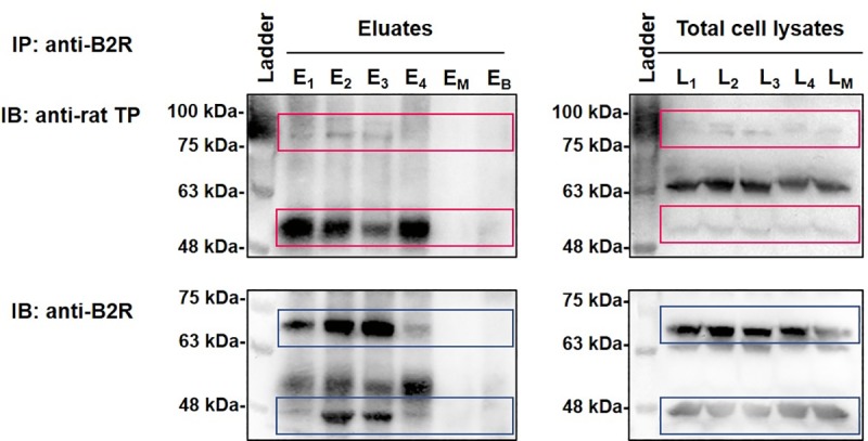
Fig 5. B2R-TP interactions in RASMC as revealed by co-IP followed by SDS PAGE.

RASMC lysates were immunoprecipitated with anti-B2R followed by immunoblotting with anti-TP (upper panels) and anti-B2R (lower panels) antibodies, successively. E and L represent eluates and matching lysates of unstimulated RASMC (E1, L1), or RASMC that were stimulated with BK 10−11 M (E2, L2), IBOP 10−7 M (E3, L3), or [BK 10−11 M + IBOP 10−7 M] (E4, L4) for 10 min. EM and LM represent eluates and matching lysates of mock co-IP condition. (EB) represents eluates of a control co-IP condition, whereby Dynabeads-protein-A-anti-B2R were incubated with PBS instead of RASMC lysates. Images are representative of three qualitatively similar independent experiments. A denatured broad molecular weight protein ladder was loaded in parallel (upper and lower left-hand lanes).
