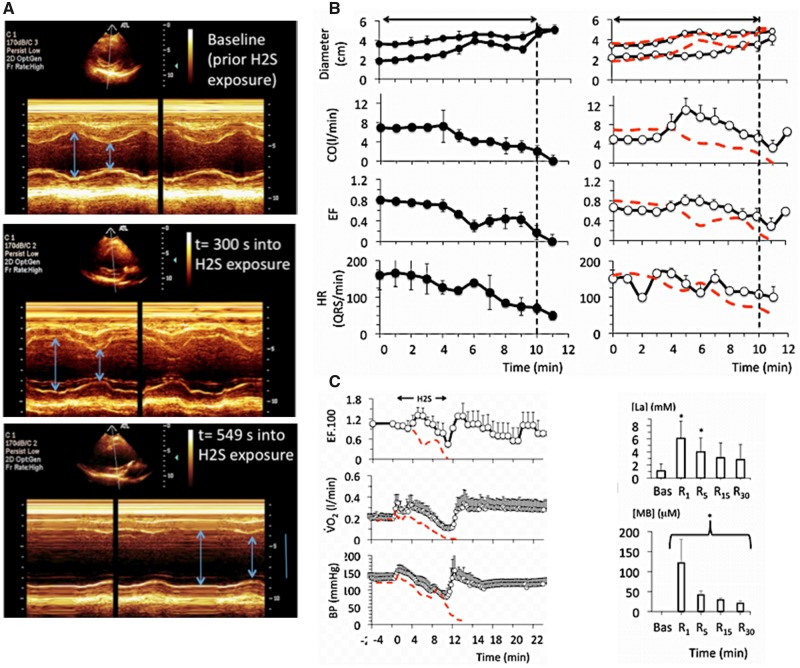
Figure 2.
A, Example in 1 sheep of a 2D echocardiography study during a lethal sulfide infusion. Note the very rapid distension of the heart and increase in left ventricular volume after few minutes of H2S exposure, leading to a suppression of the cardiac systole (PEA) in less than 10 min. B, Effects of lethal levels of H2S (protocol 1) on cardiac function determined by echocardiography in 4 untreated animals (saline, left panels), and in 4 animals treated with MB (right panels). From top to bottom, mean ± SD values of end-diastolic and end-systolic diameter, cardiac output (CO), left ventricular ejection fraction (LVEF), and heart rate (determined from the ECG signal) are displayed. In the nontreated intoxicated group, note that a rapid depression in cardiac contractility led to a cardiogenic shock followed by a cardiac asystole. Note that when cardiac contractions ceased, heart rate (ECG signal) is maintained at an average of 50 QRS/min, defining a PEA. MB administered during H2S infusion maintained the cardiac function within a range compatible with survival; for instance, cardiac output still averaged 3 l/min at a time when all the control sheep where in cardiac asystole. C, Temporal profile of the changes (mean ± SD) in LVEF), O2, and blood pressure following sulfide exposure (protocol 1, lethal administration) in the MB-treated sheep (open symbols), whereas the red-doted lines correspond to the average data obtained in the untreated animals. Time zero is the time of exposure to sulfide. Note that in the MB-treated group, all the parameters retuned to baseline within a few minutes after the cessation of the exposure to sulfide and remained stable, at a time when all the nontreated animals had already died. Lactate also returned progressively to normal reaching 2.8 mM at 30 min into recovery. Concentrations of MB remained present in the blood, but decreased form 121 μM at 1 min to 20 μM at 30 min.

