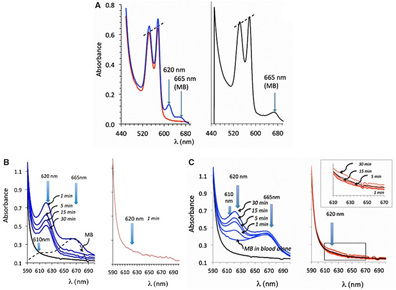
Figure 3.
A and B, Absorbance of the solution of hemoglobin obtained from the blood sampled 1 min after the end of exposure in a sheep that received H2S only (red lines, before cardiac asystole occurred) and in a sheep that received H2S plus MB (blue line). Note that a peak of absorbance at 620 nm was present when both MB and H2S were administered, for comparison the spectrum of absorbance of MB in blood in also shown (black line). C, effects of mixing blood to NaHS (100 μM), MB (60 μM), and NaHS (60 μM) plus MB (60 μM). Note that only in the latter was a peak of absorbance produced at 620 nm.

