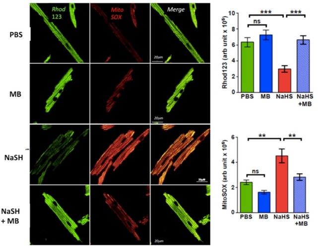
Figure 9.
Isolated LV myocytes from adult C57BL6 mice loaded with mitochondrial indicator rhodamine 123 (123 μM, Invitrogen) and mitochondrial superoxide-sensitive fluorophore MitoSOX red (22 μM; Invitrogen). Cells were exposed for 10 min to PBS, NaSH (100 μM), MB (20 μg/ml), and NaHS + MB (MB added 3 min after NaHS). Cells were imaged using a Carl Zeiss Meta 510 Meta confocal microscope was used with 1.7× digital zoom at 488 and 561 nm for rhodamine 123 and MitoSOX Red, respectively. H2S decreased the mitochondrial proton gradient, a marker of an impaired electron transport chain activity, in intoxicated cells and increased the production of reactive O2 species levels. MB rescued both responses. **p < .01; ***p < .001. Note that MB alone had no effect on the either signal.

