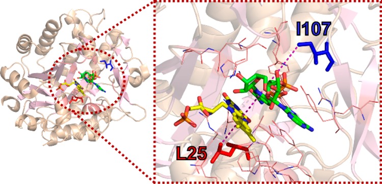
Figure 1.

Crystal structure of the PETNR−NADH4 complex (PDB: 3KFT),43 showing the location of residues L25 (red) and I107 (blue), which have been targeted for mutagenesis. Residues located 5 Å away from the NADH4 C4-H are represented as wireframes in the right panel of the figure. Residue L25 is located ∼ 4 Å away from the FMN cofactor (yellow), with the side chain pointing directly below the H-transfer coordinate, while I107 is located ∼8 Å above the coenzyme site (green) and is positioned above two bulky side chains (Y68 and Y186) that form one side of the active site hydrophobic cavity.
