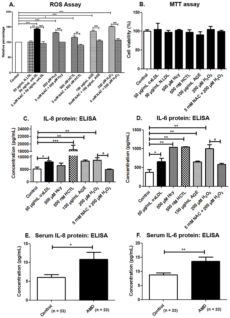
Fig 1. ROS assay and pro-inflammatory cytokine levels.
(A) ROS assay in ARPE-19 cells by DCFH-DA method, (B) MTT assay for cell viability in ARPE-19 cells, (C) IL-8 and (D) IL-6 estimation in basal conditioned media of ARPE-19 cells exposed to pro-oxidants conditions for 24 h by ELISA, (E) Serum IL-8 levels and (F) Serum IL-6 levels. Values are expressed as Mean ± SEM. *;#p < 0.05, **,##p < 0.01, ***;###;δδδp < 0.001, considered as significant. *Control vs pro-oxidants, #pro-oxidants vs NAC, δoxLDL vs N.LDL.

