. 2019 May 14;8:e42823. doi: 10.7554/eLife.42823

Figure 3. Simultaneous visualization of all six C. elegans chromosomes.

(A) Strategy to detect six chromosomes is shown. Detection probes labeled with Alexa488 (Green), Cy3 (Red), and Alexa647 (Blue), or combinations of these three fluorophores were used to label each of the six C. elegans chromosomes a different color. (B–F) Adult C. elegans were fixed and subjected to three step hybridization to detect Chromosomes 1, 2, 3, 4, 5, and X. (B) 3D maximum projection of an oocyte. Each bivalent is labeled a different color. (C) 3D maximum projection of the pachytene region of an adult germline. A magnification of one of these nuclei is shown to the right. (D) 3D maximum projections of an intestinal nucleus, (E) hypodermal nucleus, and (F) nuclei whose size and position within the animal suggest the cell is a ventral cord neuron. All images are representative of at least three independent animals.

Figure 3.

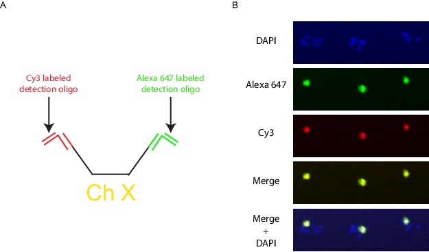
Figure 3—figure supplement 1. Multicolor labeling of the X chromosome to expand the number of detectable objects.

Figure 3—figure supplement 1.

(A) Schematic diagram of a multicolor labeling strategy. Different bridge and detection oligos are targeted to the 5’ and 3’ chromosome barcodes so that each X chromosome Oligopaint probe is targeted by both Cy3 and Alexa 647 generating an intermediate third color when the two channels are overlaid. (B) Wild-type animals were hybridized with primary probes targeting the X chromosome, and subjected to the bridge and detection oligos described in A. Signals were detected for both the Alexa 647 (green) and Cy3 (red), with the overlap generating yellow. All images are representative of at least three independent animals.
Figure 3—video 1. Six Chromosome FISH of a C. elegans intestinal nucleus.
Download video file (757.2KB, mp4)
DOI: 10.7554/eLife.42823.011
3D render of a single C. elegans intestinal nucleus from a worm hybridized with the six chromosome Oligopaint strategy. Individual chromosome territories can be clearly distinguished.