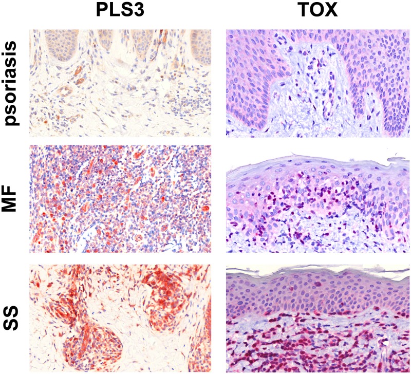
Figure 2. Immunohistochemical staining for PLS3 and TOX.
IHC staining for PLS3 of skin specimens from patients with psoriasis, MF, and SS shows strong cytoplasmic expression of PLS3 in CTCL infiltrate but not psoriasis. In all three samples, vessels are positive due to the presence of actin-bound PLS3 [35]. IHC staining for TOX shows strong, nonuniform nuclear staining in CTCL infiltrate but not psoriasis. All images are 40X and were captured using a Zeiss Mirax Midi Slide Scanner with Pannoramic MIDI/Viewer software.

