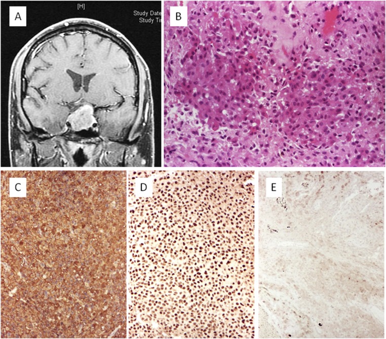
Figure 6.
Silent corticotroph adenoma: noninvasive macroadenoma. (A) Axial contrast-enhanced T1-weighted sequence. (B) Histologic slide showing sheets and acini with uniform, medium-size cells with basophilic cytoplasm (hematoxylin and eosin stain, ×20). (C) Expression of ACTH is diffuse (immunoperoxidase stain, ×10). (D) Neoplastic cells show nuclear expression of the transcription factor TPIT (immunoperoxidase stain, ×10). (E) No expression of PC1/3 was present in the tumor cells (immunoperoxidase stain, ×10).

