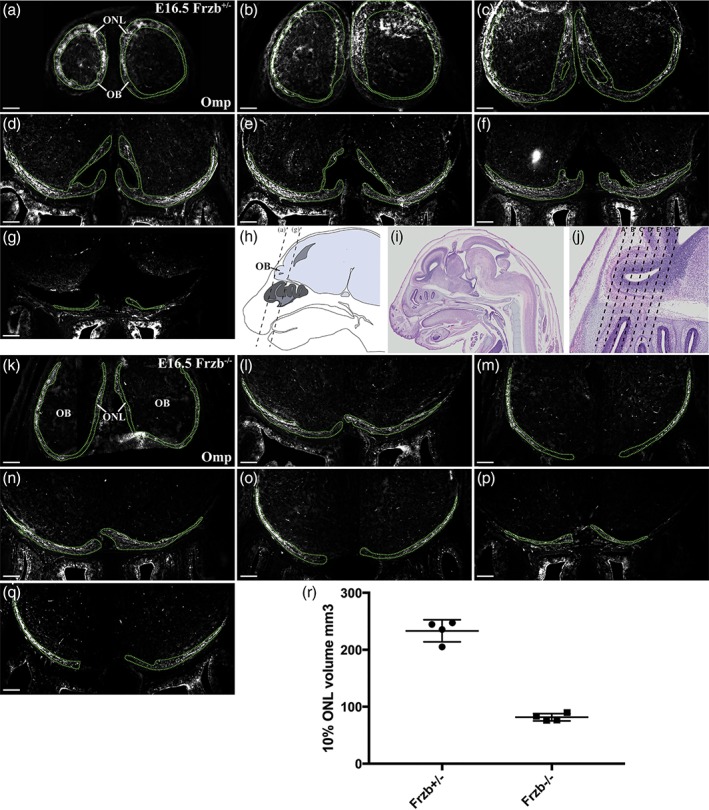
Figure 5.

The volume of the Omp‐immunoreactive olfactory nerve layer is reduced after Frzb deletion. (a–g) Coronal sections moving rostrally to caudally through the olfactory bulbs of a heterozygous Frzb +/− mouse embryo, immunostained for Omp, with the Omp‐positive regions outlined. (h) Schematic showing a parasagittal view through an E16.5 mouse head, with dotted lines showing the approximate location of the most rostral and most caudal sections shown in (a–g). (i,j) Low‐power (i) and high‐power (j) views of a hematoxylin and eosin‐stained parasagittal section through an E16.5 mouse head (Plate 38c, image a, from the eHistology Atlas with Kaufman annotations; Graham et al., 2015). The dotted lines in (j) show the approximate locations of the sections shown in (a–g). (k–q) Coronal sections moving rostrally to caudally through the olfactory bulbs of a Frzb‐null mouse embryo, immunostained for Omp, with the Omp‐positive regions outlined. (r) Scatter plot showing the mean volume of one‐tenth of the Omp‐positive ONL at E16.5 in heterozygous Frzb +/− embryos (n = 4) and Frzb‐null embryos (n = 5). Error bars show SD. Scale bar: 200 μm [Color figure can be viewed at wileyonlinelibrary.com]
