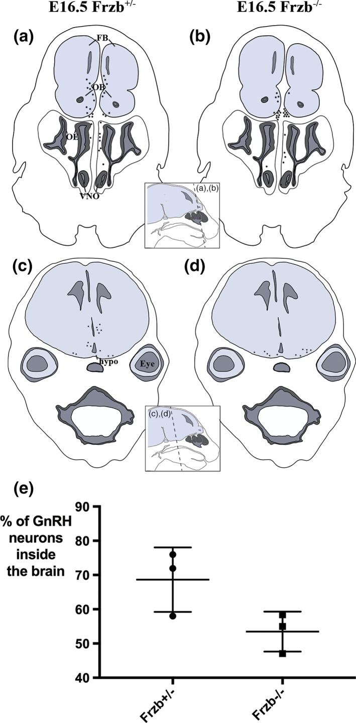
Figure 7.

Frzb deletion does not significantly affect GnRH neuron entry into the forebrain. (a–d) Schematic representations of the distribution of GnRH neurons (black spots) at E16.5 on coronal sections at different rostrocaudal levels of a heterozygous Frzb +/− mouse embryo (a,b) and a Frzb‐null mouse embryo (c,d). Each dot represents a single GnRH neuron counted on one slide of a 10‐slide series. (e) Scatter plot showing the mean percentage of GnRH neurons found inside the brain for heterozygous Frzb +/− embryos (n = 3) versus Frzb‐null embryos (n = 3). Error bars show SD. FB, forebrain; hypo, hypothalamus; OB, olfactory bulb, OE, olfactory epithelium; VNO, vomeronasal organ [Color figure can be viewed at wileyonlinelibrary.com]
