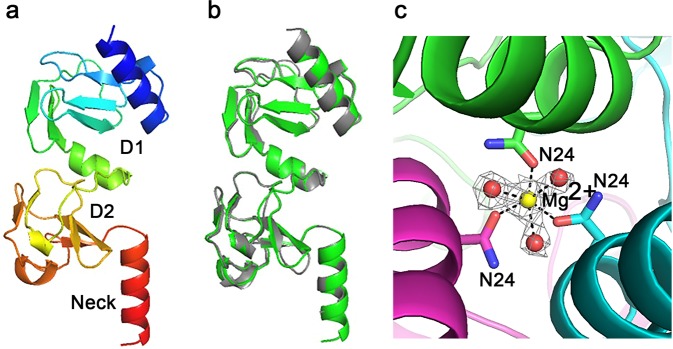
Figure 3.
TSP3 head domain structure. (a) Cartoon representation of the two N-terminal subdomains and the neck with rainbow coloring from the N-terminus (blue) to the C-terminus (red). (b) Cartoon image of superimposed head and neck regions of TSP1 (gray) and TSP3 (green). (c) Mg2+ binds along the trimer 3-fold axis close to the C-terminus of the N-terminal 3-helix bundle with octahedral coordination to an Asn24 side chain from each subunit and three water molecules. The Mg2+-ligand distances vary between 2.1 and 2.2 Å. The electron density associated with the cation and coordinating water molecules is shown (2Fo-Fc) coefficients and 1σ level. Carbon atoms and secondary structure unit of each subunits are shown in different colors. Oxygens and nitrogen atoms are colored red and blue, respectively.

