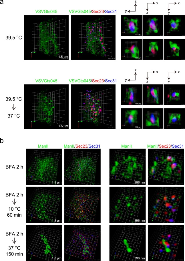
Figure 2.
Cargoes are concentrated into domains within the ER exit sites for secretion. (a) HeLa cells were transfected with VSVG-ts045-GFP. The cells were cultured at 39.5 °C to accumulate the protein within the ER, and fixed (upper panel), or further incubated at 37 °C for 8 min before fixation (bottom panel). The cells were stained with anti-Sec23 (red) and anti-Sec31 (blue) antibodies. 3D triple-color observation by SCLIM is shown. Right, magnifications of images on the left with 2D projection. The length indicates the scale of each unit. (b) HeLa cells stably expressing ManII-GFP were incubated with 5 µg/ml brefeldinA for 2 h, washed with ice-cold DMEM supplemented with 10% fetal bovine serum, and incubated either at 10 °C for 60 min or at 37 °C for 150 min before fixation. Fixed cells were processed for immunofluorescence. The cells were stained with anti-Sec23 (red) and anti-Sec31 (blue) antibodies. 3D triple-color observation by SCLIM is shown. Right, magnifications of images on the left. The length indicates the scale of each unit.

