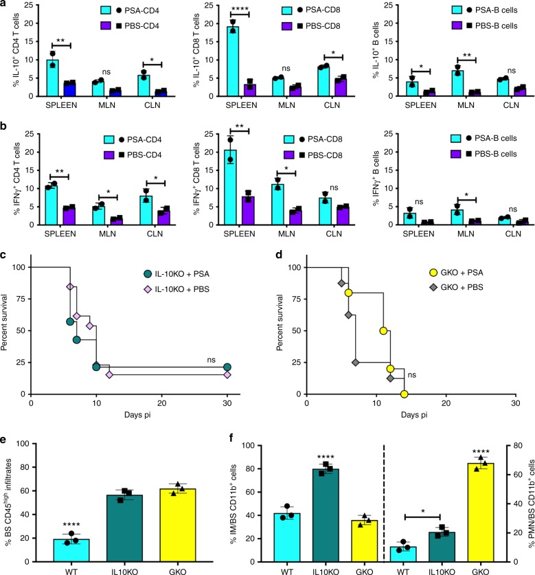
Fig. 4.
PSA increases IL-10 and IFNγ-secreting T cells. CD4 and CD8 T cells and B cells in spleens, mesenteric lymph nodes (MLN), and cervical lymph nodes (CLN) of PSA or PBS-treated WT mice at day 6 pi were analyzed for a IL-10 and b IFNγ secretion, n = 2 experiments; *p < 0.05, **p < 0.01, ****p < 0.0001, as determined by two-way ANOVA with Sidak’s multiple comparisons test. Survival of PSA or PBS treated c IL-10KO mice or d IFN-GKO mice (n = 8–16 mice); ns: not significant. Bar plots show e % CD45high leukocytes, f (left y-axis) % Ly6Chigh IM and (right y-axis) % Ly6G+ neutrophils (PMN) within CD45high CD11b+ cells infiltrating the BS of PSA treated 129 WT, IL10KO, and GKO mice at day 6 pi, n = 3 experiments with 2–3 BS/group; *p < 0.05, ****p < 0.0001 as determined by ordinary one-way ANOVA with Turkey’s multiple comparisons tests

