Figure 1.
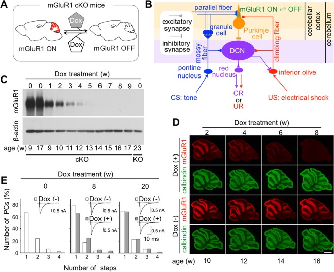
Climbing fiber innervation of PCs is normal after mGluR1 depletion in adult mGluR1 cKO mice. (A) mGluR1 in mGluR1 cKO PCs is reversibly inactivated by Dox administration. (B) Simplified schematic diagram of the circuits essential for eyeblink conditioning. Most interneurons have been omitted from this diagram. (C) Immunoblotting of cerebellar proteins from Dox-treated and untreated cKO mice with anti-mGluR1 and anti-β-actin antibodies. All lanes contain 10 µg of protein. The lane at the right end contains proteins from a global mGluR1 KO mouse16. (D) Immunostaining of cerebellar sagittal slices from Dox-treated and untreated cKO mice with anti-mGluR1 (red) and calbindin (green) antibodies. Scale bar: 1 mm. (E) Summary histograms showing the number of discrete steps for climbing fiber-mediated EPSCs (CF-EPSCs) recorded from mGluR1 cKO PCs before (left, 45 cells), 8 weeks after [middle, 56 cells for Dox (−) and 55 cells for Dox (+)], and 20 weeks after [right, 30 cells for Dox (−) and 57 cells for Dox (+)] Dox treatment. No significant difference was found between Dox-treated and untreated mice after 8 weeks (p = 0.110, Mann-Whitney U-test) or 20 weeks (p = 0. 839). Insets show representative traces of CF-EPSCs recorded from PCs.

