Figure 2.
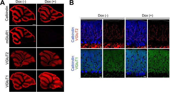
Normal distribution of parallel fiber and climbing fiber terminals 37 weeks after the initiation of Dox administration. (A) Immunofluorescence of cerebellar parasagittal sections from untreated and Dox-treated cKO mice 37 weeks after the beginning of Dox administration. Sections were stained with antibodies against mGluR1, calbindin, VGluT1 and VGluT2. In Dox-treated cKO mice, immunoreactivity for mGluR1 was abolished, whereas that for calbindin, VGluT1, or VGluT2 was comparable to untreated mGluR1 cKO mice. Scale bar: 0.5 mm. (B) Triple immunofluorescence for calbindin (blue), VGluT1 (green) and VGluT2 (red). There was no apparent difference between untreated and Dox-treated mGluR1 cKO mice in the distribution of parallel fiber or climbing fiber terminals. Scale bars: 20 µm.

