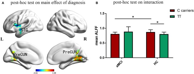
Figure 2.
Post-hoc analyses. Thresholds were set at a corrected p < 0.05, determined by Monte Carlo stimulation. (A) Compared with the control group, aMCI participants showed a significant decrease of ALFF in left inferior frontal gyrus, superior temporal gyrus and insula, while increased ALFF in right cuneus, calcarine and bilateral posterior cingulate and precuneus. (B) Further post-hoc tests of the significant interactions of diagnosis and genotype revealed that, aMCI C carriers showed decreased ALFF in the interactive brain regions compared with control C carriers. In addition, the ALFF value of C carriers was higher than that of TT genotype in control group. IFG inferior frontal gyrus, STG superior temporal gyrus, CAL calcarine, PreCUN precuneus. *Indicates a statistical difference between groups, P < 0.05.

