Figure 2.

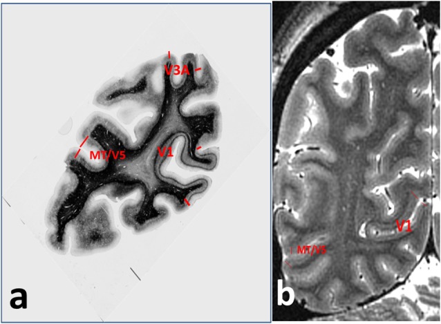
Annotations for Figure 1, showing (A) cortical areas V1, V3A and MT/V5 on the myelin-stained histological section, and (B) V1 and MT/V5 on the MRI section.

Annotations for Figure 1, showing (A) cortical areas V1, V3A and MT/V5 on the myelin-stained histological section, and (B) V1 and MT/V5 on the MRI section.