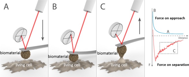
Figure 1.
Schematic representation of the measurement of cell-biomaterial interaction forces by colloidal probe microscopy. A biomaterial-coated colloidal probe and a substrate with living cells are approached each other (A) until contact (B), and then they are retracted (C) until detachment. The interaction forces are quantified from the deflection of the cantilever, which is monitored with a laser and a photodetector. Figure prepared by Joel Wolff.

